. 2011 Oct;36(9):1312-26.
doi: 10.1016/j.psyneuen.2011.03.003. Epub 2011 Apr 9.

Neuroanatomical evidence for reciprocal regulation of the corticotrophin-releasing factor and oxytocin systems in the hypothalamus and the bed nucleus of the stria terminalis of the rat: Implications for balancing stress and affect

Affiliations

Neuroanatomical evidence for reciprocal regulation of the corticotrophin-releasing factor and oxytocin systems in the hypothalamus and the bed nucleus of the stria terminalis of the rat: Implications for balancing stress and affect

Joanna Dabrowska et al. Psychoneuroendocrinology. 2011 Oct.

Abstract

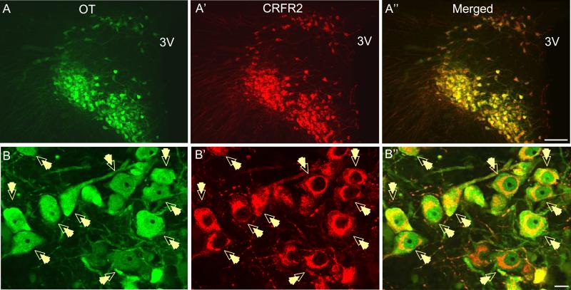
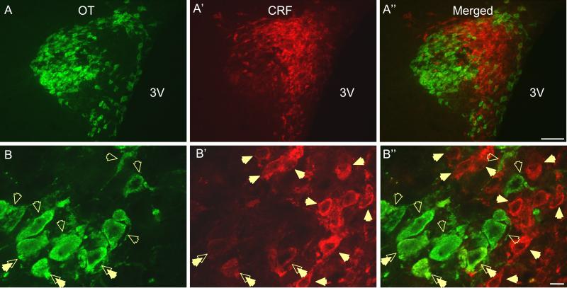
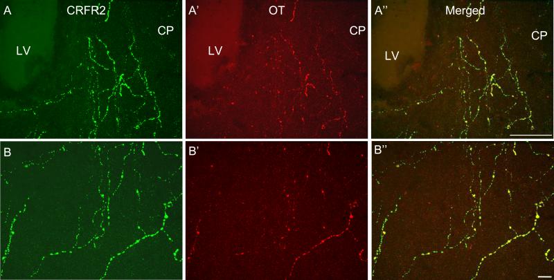
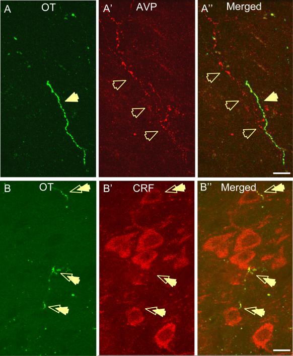
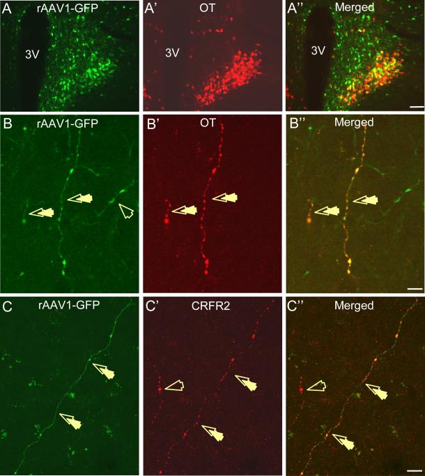
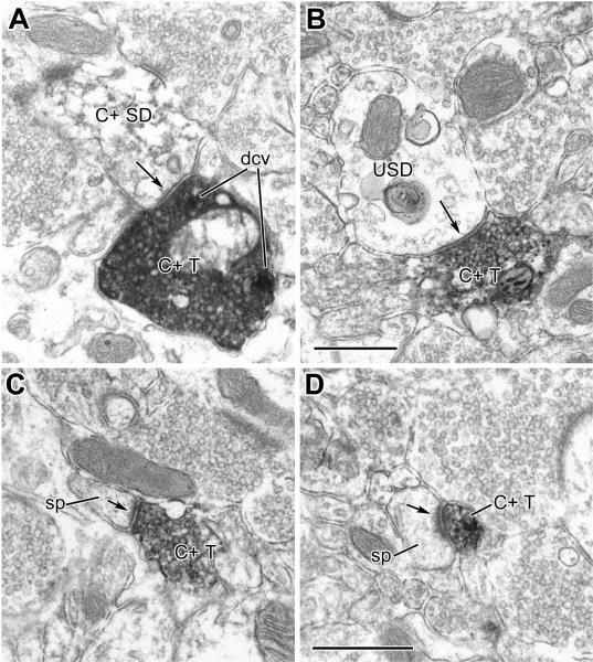
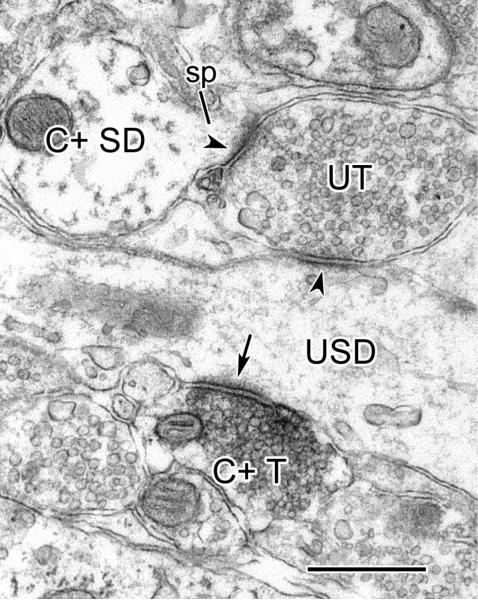
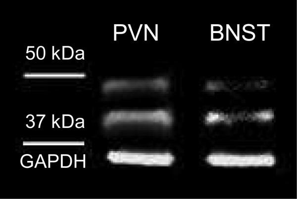
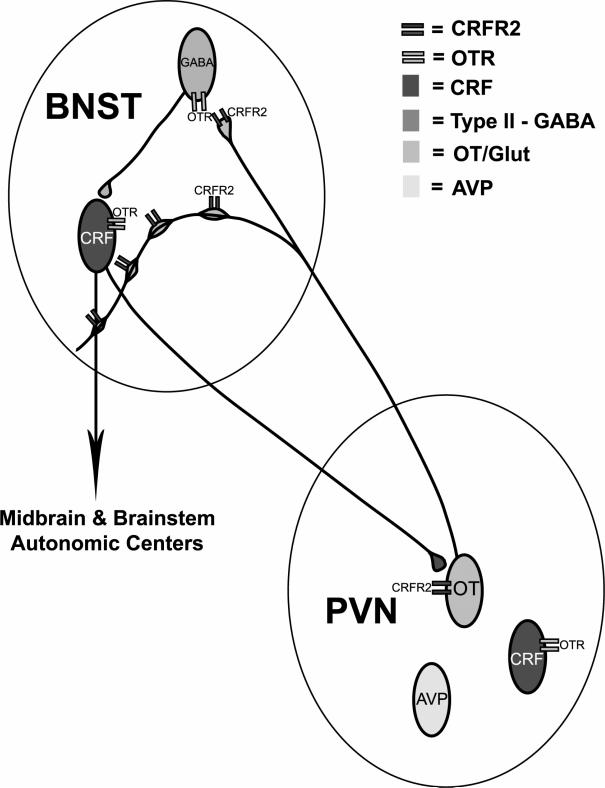
Activation of corticotrophin releasing factor (CRF) neurons in the paraventricular nucleus of the hypothalamus (PVN) is necessary for establishing the classic endocrine response to stress, while activation of forebrain CRF neurons mediates affective components of the stress response. Previous studies have reported that mRNA for CRF2 receptor (CRFR2) is expressed in the bed nucleus of the stria terminalis (BNST) as well as hypothalamic nuclei, but little is known about the localization and cellular distribution of CRFR2 in these regions. Using immunofluorescence with confocal microscopy, as well as electron microscopy, we demonstrate that in the BNST CRFR2-immunoreactive fibers represent moderate to strong labeling on axons terminals. Dual-immunofluorescence demonstrated that CRFR2-fibers co-localize oxytocin (OT), but not arginine-vasopressin (AVP), and make perisomatic contacts with CRF neurons. Dual-immunofluorescence and single cell RT-PCR demonstrate that in the hypothalamus, CRFR2 immunoreactivity and mRNA are found in OT, but not in CRF or AVP-neurons. Furthermore, CRF neurons of the PVN and BNST express mRNA for the oxytocin receptor, while the majority of OT/CRFR2 neurons in the hypothalamus do not. Finally, using adenoviral-based anterograde tracing of PVN neurons, we show that OT/CRFR2-immunoreactive fibers observed in the BNST originate in the PVN. Our results strongly suggest that CRFR2 located on oxytocinergic neurons and axon terminals might regulate the release of this neuropeptide and hence might be a crucial part of potential feedback loop between the hypothalamic oxytocin system and the forebrain CRF system that could significantly impact affective and social behaviors, in particular during times of stress.


Figures

Figure 1
Figure 1
A-A” Photomicrographs showing high level co-localization of CRFR2 (A’, red) and OT (A, green) in the PVN (A”, merged, 10x, scale bar 100 μm). Both CRFR2 (A’, red) and OT (A, green) demonstrate high somatodendritic immunoreactivity in the PVN neurons. B-B” As seen at higher magnification (63x, scale bar 10 μm), all CRFR2-immunoreactive neurons (B’, red) co-localize OT (B, green) at the level of soma, dendrites and axons in the PVN (B”, merged; 3V - 3rd ventricle).
Figure 2
Figure 2
A-A” Photomicrographs showing adjacent and mainly non-overlapping somatodendritic immunoreactivity of OT (A, green) and CRF (A’, red) in the PVN (A”, merged, magnification 10x, scale bar 100 μm, 3V – 3rd ventricle). B-B” At higher magnification (63x, scale bar 10 μm) it can be seen that CRF (B’, red, closed arrows) and OT (B, green, open arrows) represent exclusive neuronal populations in the PVN (B”, merged). Occasionally, OT-positive cells demonstrate immunolabeling for CRF as indicated by double arrows.
Figure 3
Figure 3
A-A” Photomicrographs showing high level co-localization of CRFR2-immunoreactive fibers (A, green) and OT-positive fibers (A’, red) in the anterolateral BNST group (BNSTALG, A”, 20x, scale bar 100 μm). B-B” As seen at higher magnification (63x, scale bar 10 μm), both CRFR2- (B, green) and OT- (B’, red) immunoreactive fibers are characterized by multiple, beaded varicosities, indicative of possible en passant release sites and axon terminals and the great majority of CRFR2-immunopositive fibers co-localize OT in the BNSTALG (B”, merged, LV – lateral ventricle, CP – Caudate Putamen). All of the photomicrographs shown above represent compiled Z-stack images taken at 50 μm resolution through the z-axis of the tissue and processed using Simple PCI 6.6 software.
Figure 4
Figure 4
A-A” Photomicrographs showing exclusive immunoreactivity of OT (A, green) and AVP-positive fibers (A’, red) in the BNSTALG. Both AVP and OT-immunoreactive fibers are highly expressed in the BNSTALG but they are exclusively localized with each other (A”, merged, 63x, scale bar 10 μm). B-B” Photomicrographs showing exclusive OT- (B, green) and CRF- (B’, red) immunoreactivity in the BNSTALG. High somatodendritic immunoreactivity of CRF is seen in the BNSTALG with the highest expression in the oval nucleus division. As indicated by double arrows, OT-positive fibers make perisomatic contacts with CRF-immunoreactive cells in the BNSTALG (B”, merged, 63x, scale bar 10 μm).
Figure 5
Figure 5
Figure 5. A-A” Photomicrographs showing high level co-localization of GFP (A, green) with OT (A’, red) in the PVN fourteen days after rAAV1-CMV-GFP was injected into the PVN. GFP expression was restricted to the PVN neurons and some of GFP-immunoreactive neurons co-localized OT (A”, merged) and CRFR2 (not shown, 10x, scale bar 100 μm). B-B”-C-C” Significantly, GFP-labeled fibers (B, C, green), with beaded varicosities were also observed in the BNSTALG and some of these fibers co-localized OT (B”, merged) and CRFR2 (C”, merged, double arrows, 63x, scale bar 10 μm).
Figure 6.1
Figure 6.1
Electron micrographs of CRFR2-immunoreactivity in the BNSTALG. A: A CRFR2-ir axon terminal (C+ T) makes asymmetrical synaptic contact (arrow) with a lightly-immunoreactive small caliber dendrite (C+ SD). The postsynaptic density appears thicker in serial sections. Two dense core vesicles (DCV) in the labeled terminal are indicated. B: A CRFR2-ir axon terminal (C+ T) makes symmetrical synaptic contact (arrow) with an unlabeled small caliber dendrite (USD). C, D: CRFR2-ir terminals (C+ T) make asymmetrical synaptic contacts (arrows) with dendritic spines (sp). Scale bars = 0.5 μm: Scale in B is for A and B; scale in D is for C and D.
Figure 6.2
Figure 6.2
Electron micrograph of a CRFR2-ir terminal (C+ T) making asymmetrical synaptic contact (arrow) with an unlabeled small caliber dendrite (USD) in the BNSTALG. This unlabeled dendrite also receives an asymmetrical synapse (arrowhead) from an unlabeled terminal (UT). The unlabeled terminal also makes asymmetrical synaptic contact (arrowhead) with a spine (sp) emerging from a lightly immunoreactive dendrite (C+ SD). Scale bar = 0.5 μm.
Figure 7
Figure 7
Expression of CRFR2 protein in both PVN and BNST tissue extracts was confirmed by presence of two distinct bands in the immunoblots at ~ 48 and 40 kDa, which correspond, respectively, to full-length CRFR2 protein and soluble splice variant of CRFR2.
Figure 8
Figure 8
Schematic demonstration of potential feedback loop between CRF and OT systems based on reciprocal neuroanatomical relationship between CRF-containing neurons of the BNSTALG and PVN and OT-containing neurons of the hypothalamus. OT receptors are expressed by CRF neurons of the PVN and BNSTALG and conversely CRFR2 are expressed by OT neurons in the PVN and consequently fibers in the BNSTALG.

Similar articles

Cited by

References

    1. Alheid GF. Extended amygdala and basal forebrain. Ann N Y Acad Sci. 2003;985:185–205. - PubMed
    1. Amico JA, Cai HM, Vollmer RR. Corticosterone release in oxytocin gene deletion mice following exposure to psychogenic versus non-psychogenic stress. Neurosci Lett. 2008;442:262–266. - PMC - PubMed
    1. Amico JA, Mantella RC, Vollmer RR, Li X. Anxiety and stress responses in female oxytocin deficient mice. J Neuroendocrinol. 2004;16:319–324. - PubMed
    1. Arima H, Aguilera G. Vasopressin and oxytocin neurones of hypothalamic supraoptic and paraventricular nuclei co-express mRNA for Type-1 and Type-2 corticotropin-releasing hormone receptors. J Neuroendocrinol. 2000;12:833–842. - PubMed
    1. Arluison M, Vankova M, Cesselin F, Leviel V. Origin of some enkephalin-containing afferents to the ventro-medial region of the globus pallidus in the rat. Brain Res Bull. 1990;25:25–34. - PubMed

Publication types

MeSH terms