Review
. 2011 Apr 13;19(4):447-59.
doi: 10.1016/j.str.2011.02.004.

The structural biology of Toll-like receptors

Affiliations
Review

The structural biology of Toll-like receptors

Istvan Botos et al. Structure. .

Abstract

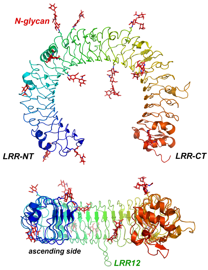
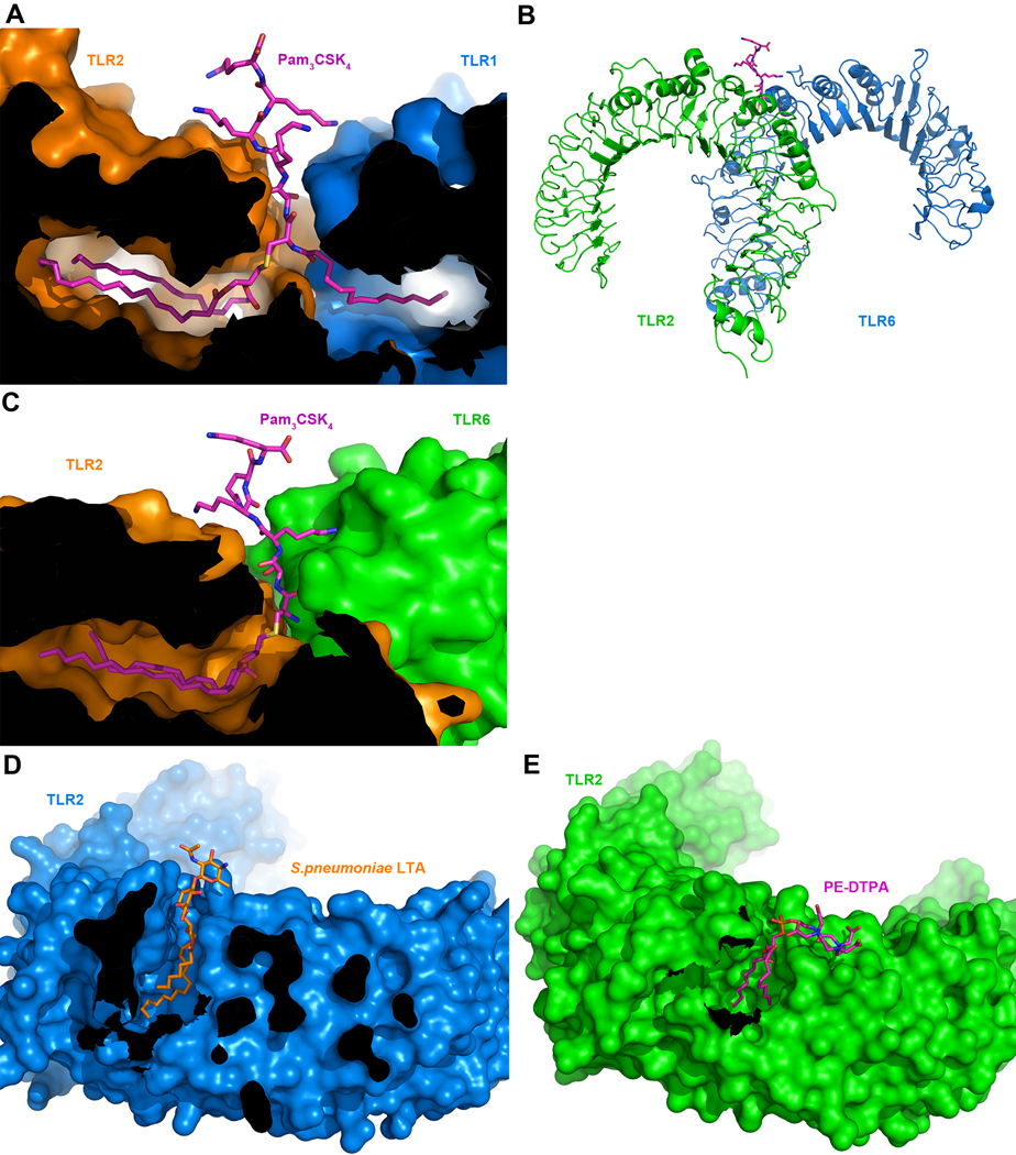
The membrane-bound Toll-like receptors (TLRs) trigger innate immune responses after recognition of a wide variety of pathogen-derived compounds. Despite the wide range of ligands recognized by TLRs, the receptors share a common structural framework in their extracellular, ligand-binding domains. These domains all adopt horseshoe-shaped structures built from leucine-rich repeat motifs. Typically, on ligand binding, two extracellular domains form an "m"-shaped dimer sandwiching the ligand molecule bringing the transmembrane and cytoplasmic domains in close proximity and triggering a downstream signaling cascade. Although the ligand-induced dimerization of these receptors has many common features, the nature of the interactions of the TLR extracellular domains with their ligands varies markedly between TLR paralogs.


Figures

Figure 1
Figure 1
The structure of Leucine-Rich Repeats. (A) LRR consensus sequences for TLR3 and Ribonuclease Inhibitor. Residues forming the β strand are highlighted in orange. (B) A LRR loop from hTLR3 and a LRR loop from RI, with the conserved residues forming a hydrophobic core. The boxed regions form the surfaces involved in ligand binding. (C) Ribbon diagram of TLR3 (Bell et al., 2005) (2A0Z) and Ribonuclease Inhibitor (Kobe et al., 1995) (1DFJ). (D) Ribonuclease inhibitor complexed with ribonuclease A (Kobe et al., 1995) (1DFJ). The LRR protein is shown in blue and the ligand in orange. (E) Lamprey VLR complexed with H-trisaccharide (Han et al., 2008) (3E6J). (F) Glycoprotein Ib alpha complexed with the von Willebrand factor A1 domain (Huizinga et al., 2002) (1M10). Figures generated with program Pymol (DeLano, 2002).
Figure 2
Figure 2
The structure of a TLR-ECD (hTLR3). Top and side views of the TLR3-ECD, with the N-linked glycosyl moieties (Bell et al., 2005) (2A0Z). The LRRs are capped by the LRR-NT and LRR-CT motifs and the molecule is flat with one glycan-free side (ascending side) that is involved in receptor dimerization.
Figure 3
Figure 3
Structure of the TLR1/TLR2/Pam3CSK4 complex. (A) The TLR2-ECD showing the position of the three subdomains: N-terminal, central and C-terminal. (B) hTLR1 and Pam3CSK4 ligand interactions mapped on the molecular surface. (C) hTLR2 and Pam3CSK4 interactions mapped on the surface. (D) Ribbon diagram of TLR1/TLR2 binding Pam3CSK4 (magenta) (Jin et al., 2007) (2Z7X).
Figure 4
Figure 4
Ligand binding by TLR 1, 2 and 6. (A) Cross-section through the molecular surface of the TLR1/TLR2/Pam3CSK4 complex (2Z7X). (B) Ribbon diagram of TLR2/TLR6 binding Pam2CSK4 (magenta) (Kang et al., 2009)(3A79). (C) Cross-section through the molecular surface of the TLR2/TLR6/Pam2CSK4 complex (3A79). (D) Cross-section through the molecular surface of the TLR2/pnLTA complex (Kang et al., 2009)(3A7B). (E) Cross-section through the molecular surface of the TLR2/PE-DTPA complex (Kang et al., 2009)(3A7C).
Figure 5
Figure 5
Structure of the TLR3/dsRNA complex. (A) Molecular surface of TLR3 dimer (green) with bound dsRNA (Liu et al., 2008)(3CIY). The interaction of the C-terminal capping motifs stabilizes the TLR3 dimer. (B) Top view.
Figure 6
Figure 6
Structure of the TLR4/MD-2/LPS complex. (A) Ribbon diagram of TLR4 green, MD-2 (blue) binding LPS (magenta) (Park et al., 2009)(3FXI). (B) Cross-section through the molecular surface of the MD-2/LPS complex. (C) TLR4 and LPS interactions mapped on their molecular surface. (D) Cross-section through the molecular surface of the MD-2/lipid IVa complex (Ohto et al., 2007)(2E59). (E) Cross-section through the molecular surface of the MD-2/Eritoran complex (Kim et al., 2007) (2Z65).
Figure 7
Figure 7
(A) Structural model of the full-length TLR3/dsRNA signaling complex. The model is based on the mTLR3/dsRNA structure (3CIY) and a TLR3 TIR domain homology model based on the TLR10 TIR structure (2J67). The transmembrane portions have been modeled as α-helices. (B) TLR signaling pathways. TLR3 signals exclusively through TRIF, while TLR4 can use both the TRIF and the MyD88 pathways. All other TLRs use the MyD88 pathway.
Figure 8
Figure 8
Structure of the TIR domain dimer from TLR10. (A) Ribbon diagram of the dimeric TIR10, with the two interacting BB-loops highlighted in gold and red (Nyman et al., 2008) (2J67). (B) Molecular surface of dimeric TIR10.

References

    1. Akashi-Takamura S, Miyake K. TLR accessory molecules. Curr. Opin. Immunol. 2008;20:420–425. - PubMed
    1. Andersen-Nissen E, Smith KD, Bonneau R, Strong RK, Aderem A. A conserved surface on Toll-like receptor 5 recognizes bacterial flagellin. J. Exp. Med. 2007;204:393–403. - PMC - PubMed
    1. Asagiri M, Hirai T, Kunigami T, Kamano S, Gober HJ, Okamoto K, Nishikawa K, Latz E, Golenbock DT, Aoki K, Ohya K, Imai Y, Morishita Y, Miyazono K, Kato S, Saftig P, Takayanagi H. Cathepsin K-dependent toll-like receptor 9 signaling revealed in experimental arthritis. Science. 2008;319:624–627. - PubMed
    1. Bell JK, Botos I, Hall PR, Askins J, Shiloach J, Segal DM, Davies DR. The molecular structure of the Toll-like receptor 3 ligand-binding domain. Proc. Natl. Acad. Sci. U. S. A. 2005;102:10976–10980. - PMC - PubMed
    1. Bell JK, Mullen GE, Leifer CA, Mazzoni A, Davies DR, Segal DM. Leucine-rich repeats and pathogen recognition in Toll-like receptors. Trends Immunol. 2003;24:528–533. - PubMed

Publication types

Substances

LinkOut - more resources