

Coagulopathy during cardiac arrest and resuscitation in a swine model of electrically induced ventricular fibrillation
- PMID: 21482008
- PMCID: PMC3549665
- DOI: 10.1016/j.resuscitation.2011.02.034
Coagulopathy during cardiac arrest and resuscitation in a swine model of electrically induced ventricular fibrillation
Abstract
Aims: Coagulopathy is often present after resuscitation from cardiac arrest but plays an undefined role in the post cardiac arrest syndrome. The aim of this study was to characterize coagulation changes during cardiac arrest and post-resuscitation care in order to direct further focused study.
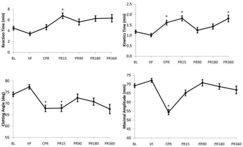
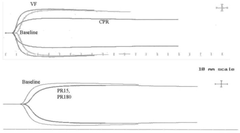
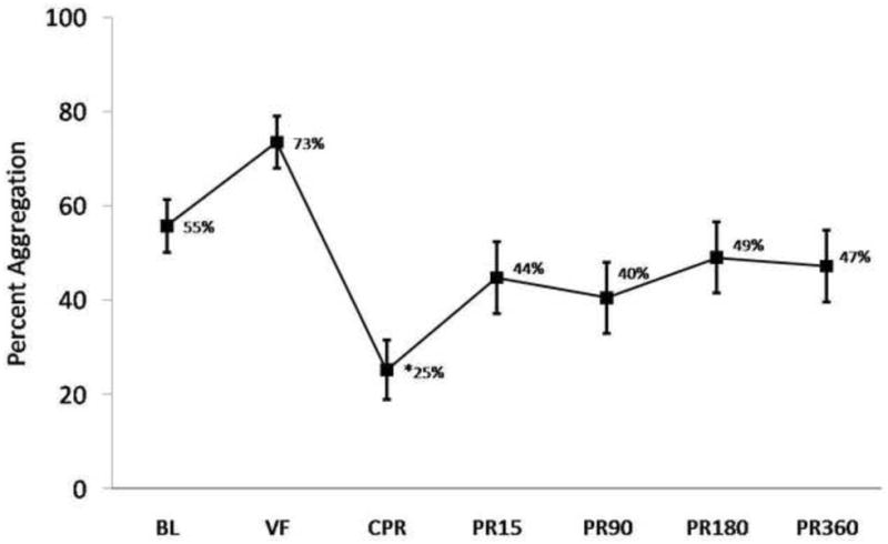
Methods: Ventricular fibrillation (VF) was induced electrically in immature male swine, followed by normothermic American Heart Association Advanced Cardiac Life Support and a uniform post-resuscitation goal-directed resuscitation protocol. PT, aPTT, fibrinogen, Thrombelastography (TEG), platelet contractile force (PCF), clot elastic modulus (CEM), and collagen-induced platelet aggregation were compared at baseline, at 8 min of VF, during the 3rd round of chest compressions (CPR), and at 15, 90, 180, and 360 min after return of circulation using repeated measures ANOVA.
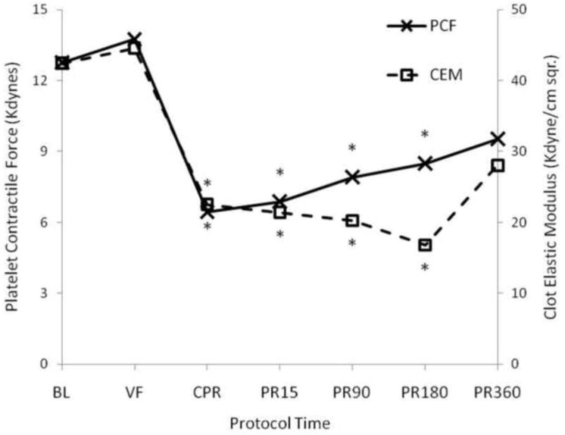
Results: 8/18 (44%) animals were resuscitated after 10.9 ± 0.9 min of VF and 7.6 ± 3.4 min of CPR. TEG revealed a significant impairment in clot strength (MA) and clot formation kinetics (K, alpha angle) arising during CPR, followed by a brief prolongation of clot onset times (R) after return of circulation. Both PCF and CEM fell significantly during CPR (PCF by 50%, CEM by 47% of baseline) and platelet aggregation was significantly decreased during CPR. Coagulation changes were partially recovered by 3h of post-resuscitation care.
Conclusion: Whole blood coagulation was rapidly impaired during CPR after electrically induced VF in this swine model by impaired platelet aggregation/contractile function and clotting kinetics. Further platelet-specific study is indicated.
Copyright © 2011 Elsevier Ireland Ltd. All rights reserved.
Conflict of interest statement
Conflict of interest statement: None to declare.
Figures
Similar articles
-
Left ventricular thrombus development during ventricular fibrillation and resolution during resuscitation in a swine model of sudden cardiac arrest.Resuscitation. 2014 May;85(5):689-93. doi: 10.1016/j.resuscitation.2014.01.030. Epub 2014 Feb 8. Resuscitation. 2014. PMID: 24518559
-
A randomized comparison of cardiocerebral and cardiopulmonary resuscitation using a swine model of prolonged ventricular fibrillation.Resuscitation. 2010 May;81(5):596-602. doi: 10.1016/j.resuscitation.2010.01.013. Epub 2010 Feb 21. Resuscitation. 2010. PMID: 20176434
-
Immediate countershock versus cardiopulmonary resuscitation before countershock in a 5-minute swine model of ventricular fibrillation arrest.Ann Emerg Med. 2000 Dec;36(6):543-6. doi: 10.1067/mem.2000.109441. Ann Emerg Med. 2000. PMID: 11097692
-
Improved neurological outcome with continuous chest compressions compared with 30:2 compressions-to-ventilations cardiopulmonary resuscitation in a realistic swine model of out-of-hospital cardiac arrest.Circulation. 2007 Nov 27;116(22):2525-30. doi: 10.1161/CIRCULATIONAHA.107.711820. Epub 2007 Nov 12. Circulation. 2007. PMID: 17998457
-
Potassium induced cardiac standstill during conventional cardiopulmonary resuscitation in a pig model of prolonged ventricular fibrillation cardiac arrest: a feasibility study.Resuscitation. 2013 Mar;84(3):378-83. doi: 10.1016/j.resuscitation.2012.08.324. Epub 2012 Aug 31. Resuscitation. 2013. PMID: 22940601
Cited by
-
Minimally invasive beating heart technique for mitral valve surgery in patients with previous sternotomy and giant left ventricle.J Cardiothorac Surg. 2020 Jun 3;15(1):122. doi: 10.1186/s13019-020-01171-6. J Cardiothorac Surg. 2020. PMID: 32493495 Free PMC article.
-
Effect of ultrasonography and fluoroscopic guidance on the incidence of complications of cannulation in extracorporeal cardiopulmonary resuscitation in out-of-hospital cardiac arrest: a retrospective observational study.BMC Anesthesiol. 2017 Jan 6;17(1):4. doi: 10.1186/s12871-016-0293-z. BMC Anesthesiol. 2017. PMID: 28125963 Free PMC article.
-
An Effective and Reproducible Model of Ventricular Fibrillation in Crossbred Yorkshire Swine (Sus scrofa) for Use in Physiologic Research.Comp Med. 2015 Oct;65(5):444-7. Comp Med. 2015. PMID: 26473349 Free PMC article.
-
Coagulopathy as a Complication of Cardiopulmonary Arrest Following Balloon Valvuloplasty in a Dog.Vet Med Sci. 2025 Jul;11(4):e70445. doi: 10.1002/vms3.70445. Vet Med Sci. 2025. PMID: 40454853 Free PMC article.
-
Thrombolytic-Enhanced Extracorporeal Cardiopulmonary Resuscitation After Prolonged Cardiac Arrest.Crit Care Med. 2016 Feb;44(2):e58-69. doi: 10.1097/CCM.0000000000001305. Crit Care Med. 2016. PMID: 26488218 Free PMC article.
References
-
- Lloyd-Jones D, Adams RJ, Brown TM, Carnethon M, Dai S, De Simone G, et al. American Heart Association Statistics Committee and Stroke Statistics Subcommittee. Executive summary: heart disease and stroke statistics--2010 update: a report from the American Heart Association. Circulation. 2010;121(7):948–54. - PubMed
-
- Nadkarni VM, Larkin GL, Peberdy MA, et al. First documented rhythm and clinical outcome from in-hospital cardiac arrest among children and adults. JAMA. 2006;295:50–7. - PubMed
-
- Nolan JP, Laver SR, Welch CA, Harrison DA, Gupta V, Rowan K. Outcome following admission to UK intensive care units after cardiac arrest: a secondary analysis of the ICNARC Case Mix Programme Database. Anaesthesia. 2007;62:1207–16. - PubMed
-
- Neumar RW, Nolan JP, Adrie C, Aibiki M, Berg RA, Bottiger BW, et al. Post-Cardiac arrest syndrome Epidemiology, pathophysiology, treatment, and prognostication. Circulation. 2008;118:2452–2483. - PubMed
Publication types
MeSH terms
Substances
Grants and funding
LinkOut - more resources
Full Text Sources
Medical
