Review
. 2011 Apr 14;70(1):8-19.
doi: 10.1016/j.neuron.2011.03.012.

Traces of Drosophila memory

Affiliations
Review

Traces of Drosophila memory

Ronald L Davis. Neuron. .

Abstract

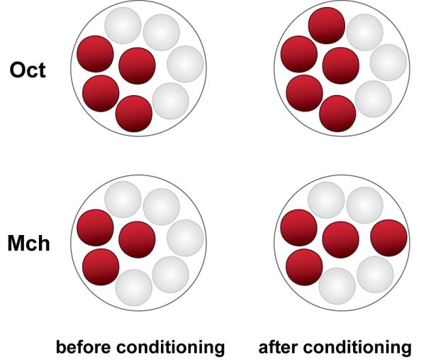
Studies using functional cellular imaging of living flies have identified six memory traces that form in the olfactory nervous system after conditioning with odors. These traces occur in distinct nodes of the olfactory nervous system, form and disappear across different windows of time, and are detected in the imaged neurons as increased calcium influx or synaptic release in response to the conditioned odor. Three traces form at or near acquisition and coexist with short-term behavioral memory. One trace forms with a delay after learning and coexists with intermediate-term behavioral memory. Two traces form many hours after acquisition and coexist with long-term behavioral memory. The transient memory traces may support behavior across the time windows of their existence. The experimental approaches for dissecting memory formation in the fly, ranging from the molecular to the systems, make it an ideal system for elucidating the logic by which the nervous system organizes and stores different temporal forms of memory.


Figures

Figure 1
Figure 1
Anatomical organization of the olfactory nervous system in Drosophila. (A) Olfactory nervous system viewed from the left-front and slightly dorsal position of the fly. Olfactory information is transmitted from olfactory receptor neurons (ORNs) located on the antennae (not shown) via the antennal nerve (AN) to the antennal lobe (AL), where the axons of ORNs synapse on two types of secondary olfactory neurons, the projection neurons (PN) and the AL interneurons (IN). The INs are known to be either excitatory or inhibitory. PNs send their axons via a nerve known as the antennal cerebral tract (ACT) to the mushroom body neurons (MBN) and to the lateral horn (LH). The PNs synapse with MBNs in a neuropil region known as the calyx (C). Three classes of MBNs have been described according to their axonal collaterals (α/β, α’/β’, and γ). The axons extended by MBNs follow the pedunculus (P) to reach the MB lobes (α, α’, β, β’, and γ). For simplicity, only one ORN axon (green), one PN (orange), one IN (purple), and one α/β MB neuron (yellow) have been superimposed on a schematic of one hemisphere of the fly brain. Axis arrows: A=anterior, D=dorsal, M=medial. Adapted from Busto et al., 2010, with permission. (B) Frontal perspective of neurons that are extrinsic to the MBs in one hemisphere showing the dorsal paired medial (DPM) neuron, anterior paired lateral (APL) neuron, and dopaminergic (DA) neurons. The DPM neuron (red) extends a single neurite which bifurcates to innervate the vertical lobes (α and α’) and the horizontal (β, β’, and γ) lobes of the MBs. Only five of the DA neurons (DA, orange) in the PPL1 cluster are illustrated. These neurons innervate distinct zones of the MB vertical lobes. The APL neuron (magenta) broadly innervates the calyx and the MB lobes. Axis arrows: D=dorsal, M=medial.
Figure 2
Figure 2
Reporter molecules used to identify memory traces in Drosophila by functional optical imaging. These reporter molecules are supplied to the organism using transgenic techniques and are expressed in a defined and limited set of neurons using the GAL4/UAS system (Brand and Perrimon, 1993). Synapto-pHluorin (spH) is a pH-sensitive GFP molecule that is trafficked to the lumen side of the synaptic vesicle by virtue of its fusion with the synaptic vesicle protein; VAMP (Miesenboeck et al., 1998). Fusion of the synaptic vesicle with the plasma membrane upon neurotransmitter release alters the pH environment around synapto-pHluorin such that its fluorescence is increased until vesicles are internalizated and reacidified. G-CaMP is a circularly permuted EGFP fused to the M13 fragment of myosin light chain kinase at one end and the calcium binding site of calmodulin on the other (Nakai et al., 2001). Upon influx of calcium through voltage-dependent calcium channels, calmodulin interacts with the M13 fragment eliciting a conformational change in EGFP that increases its fluorescence. Improved versions of G-CaMP, including G-CaMP1.6, G-CaMP2, and G-CaMP3.0, are now available (Ohkura et al., 2005; Diez-Garcia et al., 2005; Tian et al., 2009).
Figure 3
Figure 3
Schematic diagram of the experimental setup to visualize the neuronal activity in the Drosophila brain. A small window of cuticle is removed from the dorsal head capsule and the window is then sealed with plastic wrap. The living fly is mounted under the objective of a one-photon or two-photon microscope for visualizing the fluorescence of reporter molecules that are expressed from transgenes (Figure 2). Odors are puffed onto the antennae of the mounted fly from a puffer pipette (cylinder at the right of the figure) to visualize the neuronal responses to the conditioned (CS+) or unconditioned (CS-) odors for “within” animal or “between group” experiments. Electric shock is applied (for aversive learning) to the legs and abdomen of the immobilized fly with conductive wires for “within” animal experiments.
Figure 4
Figure 4
Olfactory conditioning rapidly alters the pattern of PN synaptic activity in the AL. The odorant Oct stimulates synaptic activity in four of eight dorsally located glomeruli (circles) in the AL prior to conditioning. Immediately after conditioning, five glomeruli become synaptically activated; indicating that conditioning with Oct recruits at least one additional set of PNs into its neural representation. The odor Mch also recruits an additional set of PNs into its representation after conditioning, but the set of PNs is different than that recruited by Oct. Adapted from Yu et al., 2004.
Figure 5
Figure 5
Calcium-based memory trace in the axons of the α’ neurons detected with the calcium reporter, G-CaMP. Pairing the odor of benzaldehyde (Ben) as the CS+ with mild electric shock increases the subsequent response to Ben and decreases the response to Oct (the CS-) as shown by comparing the intensity of the responses in the α’ axons before and after pairing. Note the increased number of colored pixels within the area encircled by the red dotted line (region of α’ axons) at 30 and 60 min in the Ben row compared to the “before pairing” condition, and the decreased number of colored pixels within the same area in the Oct row. No changes were detected in the axons of the α/β neurons, also shown in this image, at these timepoints after conditioning. The magnitude of response is illustrated in pseudocolor on a pixel-by-pixel basis. The α’/β’ memory trace is present at 30 min after pairing and persists for at least 60 min. Adapted from Wang et al., 2008, with permission.
Figure 6
Figure 6
Acquisition of olfactory memory in control and amn mutant flies as a function of the number of training trials. Memory increases as a function of trial number at the same rate in both control and amn mutant flies, indicating that memory formation in the mutant is essentially normal. However, memory measured at 2 hr after an equivalent amount of training is much reduced in the amn mutant flies compared to controls, indicating that the mutants are unable to consolidate their early forming memories into a stable form, or that they are impaired in an intermediate and distinct phase of memory. Adapted from Yu et al., 2005.
Figure 7
Figure 7
Intermediate-term memory trace forms in the DPM neuron processes that innervate the vertical lobes of the MBs. Grayscale images of basal fluorescence of G-CaMP expression in the distal portion of the vertical lobes from a viewpoint above the fly (see Figure 3). Calcium influx detected in the DPM processes with odor stimulation before conditioning is illustrated in the middle column of images. The magnitude of the calcium response is illustrated in pseudocolor on a pixel-by-pixel basis.The enhanced calcium influx detected in these processes with odor stimulation after conditioning is illustrated in the right column of images. Adapted from Yu et al., 2005.
Figure 8
Figure 8
A LTM trace, detected as increased calcium influx in response to the learned odor, forms in the α/β MBNs after spaced conditioning. The increased calcium influx occurs only in the vertical (α) branch of these neurons and not the horizontal (β) branch. A similar LTM trace forms in the unbranched axon of the γ MBNs after spaced conditioning but becomes detectable only by 18 hr after spaced conditioning. The α/β LTM trace is detectable by 9 hr after spaced conditioning.
Figure 9
Figure 9
Model for how memory traces may underlie temporal phases of memory after olfactory classical conditioning. Drosophila olfactory memory persists for at least 4 days after spaced conditioning and is thought to be comprised of short-term memory (STM), intermediate-term memory (ITM), long-term memory (LTM), and late-phase, long-term memory (LP-LTM). These phases of behavioral memory exist across different windows of time after conditioning, as depicted in the figure. The co-existing traces that may underlie short-term memory after conditioning include a trace that forms in the AL PNs (0-5 min), a trace in the GABAergic neuron APL (0->5 min), and a trace that forms in the α’/β’ neurons (0->1 hr). Intermediate-term behavioral memory from 30-70 min after conditioning is associated with a memory trace that forms in the DPM neurons. Long-term memory generated by spaced conditioning may be underlain by two memory cellular memory traces. A LTM trace froms in the α/β MBNs by 9 hr after spaced conditioning and persists to at least 24 hr. This memory trace is dependent on normal protein synthesis, CREB, CaMKII, the amnesiac gene product, and the gene products of 26 different genes involved in long-term behavioral memory. The most persistent memory trace discovered to date forms in the γ MBNs. This trace, which is associated with late-phase, LTM, forms by 18 hr after spaced conditioning and persists to at least 48 hr. Thus, temporal forms of behavioral memory are associated with different cellular memory traces that form in the olfactory nervous system and occupy different windows of time after conditioning.

Similar articles

Cited by

References

    1. Akalal D-BG, Wilson C, Zong L, Davis RL. Roles for Drosophila mushroom body neurons in olfactory learning. Learn. Mem. 2006;13:659–668. - PMC - PubMed
    1. Akalal D-BG, Yu D, Davis RL. A late-phase, long-term memory trace forms in the γ neurons of Drosophila mushroom bodies after olfactory classical conditioning. J Neurosci. 2010;30:16699–16708. - PMC - PubMed
    1. Akalal D-BG, Yu D, Davis RL. The long-term memory trace formed in the Drosophila α/β mushroom body neurons is abolished in long-term memory mutants. J Neurosci. 2011 in press. - PMC - PubMed
    1. Ashraf SI, McLoon AL, Sclarsic SM, Kunes S. Synaptic protein synthesis associated with memory is regulated by the RISC pathway in Drosophlia. Cell. 2006;124:191–205. - PubMed
    1. Brand AH, Perrimon N. Targeted gene expression as a means of altering cell fates and generating dominant phenotypes. Development. 1993;118:401–415. - PubMed

Publication types

LinkOut - more resources