

Review
doi: 10.1016/j.neuron.2011.03.014.
Neural systems governed by nicotinic acetylcholine receptors: emerging hypotheses
Affiliations
- PMID: 21482353
- PMCID: PMC4418790
- DOI: 10.1016/j.neuron.2011.03.014
Item in Clipboard
Review
Neural systems governed by nicotinic acetylcholine receptors: emerging hypotheses
Neuron.
.
Abstract
Cholinergic neurons and nicotinic acetylcholine receptors (nAChRs) in the brain participate in diverse functions: reward, learning and memory, mood, sensory processing, pain, and neuroprotection. Nicotinic systems also have well-known roles in drug abuse. Here, we review recent insights into nicotinic function, linking exogenous and endogenous manipulations of nAChRs to alterations in synapses, circuits, and behavior. We also discuss how these contemporary advances can motivate attempts to exploit nicotinic systems therapeutically in Parkinson's disease, cognitive decline, epilepsy, and schizophrenia.
Copyright © 2011 Elsevier Inc. All rights reserved.
Figures
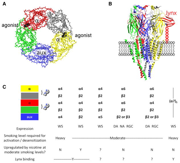
(A) A diagram of the symmetric or pseudosymmetric pentameric extracellular binding region, modeled by the acetylcholine receptor binding protein AChBP. The eyepoint is the cytosol; the side chains and transmembrane domains do not appear. The exemplar agonist (nicotine) is represented in black; two agonist binding sites form at the interface between subunits. The open state of the ion channel is more likely to occur when agonist molecules bind at both interfaces than at a single interface. An α subunit (red and yellow) always participates in the binding interface; the other participants are either α subunits (in α7 homopentameric nAChRs) or non-α subunits (in heteropentameric nAChRs such as α4β2*); (see the table in C). The auxiliary subunit (aux, in blue) does not participate in an agonist binding site. (B) Depiction of a nAChR molecule in the membrane. The eyepoint is a neighboring nAChR. The receptor is Unwin’s model for the Torpedo electric organ muscle-type AChR (Unwin, 2005). The model depicts the full extracellular region (mostly β sheets), which strongly resembles the AChBP structure shown in (A). Ribbons depict the structural elements, whereas neither backbone nor side-chain atoms appear. The model includes the full transmembrane region (mostly α-helical) and only part of the intracellular domains. The schematic also imagines a lynx molecule (red) bound at an α/non-α interface, positioned as in structures of snake α-toxins bound to AChBP (Hansen et al., 2005) or to the muscle nAChR (Dellisanti et al., 2007). Lynx binding, as independently proposed in a recent study (Lyukmanova et al., 2011), occurs at the agonist site shown in (A). The lynx molecule, unlike toxins, is tethered to the membrane by a GPI linkage, here stretched to nearly its full extent and depicted as five hexagons. (C) Some major nAChR subtypes found in brain. Each column represents the composition of a single pentameric receptor. The table shows our best present knowledge about the properties of detailed stoichiometries. The colored boxes correspond to the subunits of (A) and (B). The bracket and the nicotine molecules show the agonist-binding interfaces between individual subunits. Expression of each receptor subtype is wide-spread (WS), or restricted in the case of α6* nAChRs, confined largely to dopaminergic neurons (DA), noradrenergic neurons (NA), or retinal ganglion cells (RGC).
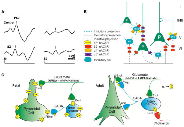
(A) Sensory inhibition deficits in schizophrenia. Cerebral evoked P50 potentials to repeated sounds (S1, S2) are inhibited in a normal (control, upper trace) but not in a schizophrenia patient (SZ, bottom trace). (B) Differential localization of nAChRs subtypes on neurons in the prefrontal cortex. Green cells are excitatory pyramidal neurons (P) and blue cells are inhibitory interneurons. FS, fast-spiking interneurons; LTS, low threshold spiking; RSNP, regular spiking nonpyramidal neuron. Adapted with permission from Poorthuis et al. (2009). (C) Development of α7nAChRs in hippocampus. In the fetal brain before cholinergic innervation occurs (left), α7 nAChRs are somatodendritic and presynaptic on both GABAergic and glutamatergic neurons. In adults (right), α7 nAChR expression is generally reduced. Receptors are still expressed on GABAergic and glutamatergic presynaptic terminals, but only GABAergic neurons express somatodendritic α7 nAChRs (figure courtesy of William Proctor).
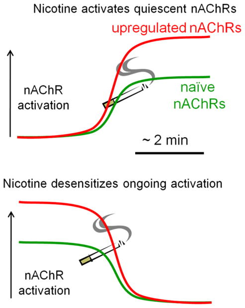
The vertical black arrow represents the level of nAChR activation at a synapse, and the x axis represents the time course of activation and/or desensitization. The cigarette represents an acute exposure to nicotine, in the context of either nicotine-naive nAChRs (green) or nicotine-upregulated receptors (red). (Top) Exposure to nicotine produces stronger activation at upregulated receptors than at naive nAChRs, because upregulated nAChRs are both more numerous and more sensitive. (Bottom) A synapse where ongoing endogenous ACh mediates stronger nAChR activation than at a naive synapse. Desensitization then produces a correspondingly larger decrement of activity. The most common example of such a desensitizing response to nicotine occurs at the presynaptic terminals of nigrostriatal dopaminergic neurons (Xiao et al., 2009).
References
-
- Adams CE. Comparison of α7 nicotinic acetylcholine receptor development in the hippocampal formation of C3H and DBA/2 mice. Brain Res Dev Brain Res. 2003;143:137–149. - PubMed
-
- Adler LE, Pachtman E, Franks RD, Pecevich M, Waldo MC, Freedman R. Neurophysiological evidence for a defect in neuronal mechanisms involved in sensory gating in schizophrenia. Biol Psychiatry. 1982;17:639–654. - PubMed
-
- Adler LE, Hoffer LD, Wiser A, Freedman R. Normalization of auditory physiology by cigarette smoking in schizophrenic patients. Am J Psychiatry. 1993;150:1856–1861. - PubMed
-
- Alkondon M, Pereira EF, Barbosa CT, Albuquerque EX. Neuronal nicotinic acetylcholine receptor activation modulates γ-aminobutyric acid release from CA1 neurons of rat hippocampal slices. J Pharmacol Exp Ther. 1997;283:1396–1411. - PubMed
Publication types
MeSH terms
Substances
Grants and funding
LinkOut - more resources
Full Text Sources
Molecular Biology Databases
