Comparative Study
. 2011 Apr 14;70(1):35-42.
doi: 10.1016/j.neuron.2011.03.001.

Experience-dependent retinogeniculate synapse remodeling is abnormal in MeCP2-deficient mice

Affiliations
Comparative Study

Experience-dependent retinogeniculate synapse remodeling is abnormal in MeCP2-deficient mice

Joao Noutel et al. Neuron. .

Abstract

Mutations in MECP2 underlie the neurodevelopmental disorder Rett syndrome (RTT). One hallmark of RTT is relatively normal development followed by a later onset of symptoms. Growing evidence suggests an etiology of disrupted synaptic function, yet it is unclear how these abnormalities explain the clinical presentation of RTT. Here we investigate synapse maturation in Mecp2-deficient mice at a circuit with distinct developmental phases: the retinogeniculate synapse. We find that synapse development in mutants is comparable to that of wild-type littermates between postnatal days 9 and 21, indicating that initial phases of synapse formation, elimination, and strengthening are not significantly affected by MeCP2 absence. However, during the subsequent experience-dependent phase of synapse remodeling, the circuit becomes abnormal in mutants as retinal innervation of relay neurons increases and retinal inputs fail to strengthen further. Moreover, synaptic plasticity in response to visual deprivation is disrupted in mutants. These results suggest a crucial role for Mecp2 in experience-dependent refinement of synaptic circuits.


Figures

Figure 1
Figure 1. Comparison of Synaptic Remodeling in −/y and +/y mice
(Left) Superimposed traces of EPSCs evoked by optic tract stimulation at increasing intensities while alternating the holding potential (HP) between −70 mV (inward currents) and +40 mV (outward currents) from +/y (P30, top) and −/y (P28, bottom) mice. (Right) The peaks of the inward (white circles) and outward (black circles) currents (through AMPA and NMDA receptors, respectively) are plotted as a function of stimulus intensity on a log scale. Grey arrows: baseline failures; white arrows: SF responses; black arrows: maximal currents (see supplemental information).
Figure 2
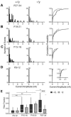
Figure 2. Sensitive Period Synaptic Remodeling is Impaired in Mecp2 −/y mice
(A–D) SF AMPAR EPSC amplitudes (HP=−70) recorded from a population of relay neurons from +/y and −/y mice over development. Amplitude histograms in 100 pA bins (left, middle panels) and cumulative probability plots (right) for ages P27–34 (A; n= 42 & 29 SF inputs for +/y and −/y, respectively), P19–21 (B; n=26 & 23), P15–16 (C; n=30 & 28) and P9–12 (D; 20 & 17). Inset in P9–12 cumulative plot shows expanded scale of the distribution. Numbers above first bin indicate total points less than 100 pA (off scale). This number includes silent inputs (SF responses with 0 AMPAR current but a measurable NMDAR current (Chen and Regehr, 2000)). The ratio of silent inputs/total inputs for P15–16 was (4/28 vs 2/30) for −/y and +/y, respectively, P19–21: (1/23 vs 0/26), and P27–34: (2/29 and 0/43). (E) Estimation of synaptic connectivity by FF for WT and −/y mice over development. For (A–E) +/y: (P9–12) 20 cells from 11 mice; (P15–16) 23 from 6; (P19–21) 22 from 14; (P27–32) 28 from 8; −/y: (P9–12) 17 from 9; (P15–16) 22 from 7; (P19–21) 17 from 10; (P27–32) 20 from 12.
Figure 3
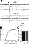
Figure 3. Comparison of Synaptic Properties in +/y and −/y mice
(A) Representative recordings of evoked quantal events from P27–34 animals in a saline solution containing 3 mM SrCl2 and 2 mM MgCl2. Arrowhead indicates time of optic tract stimulation. Stimulus artifact blanked for clarity. (B) Thousands of quantal events from many experiments were used to build cumulative probability distributions of quantal amplitudes (+/y: n=1097 events from 5 cells; −/y: n=1010 from 3 cells). The median mEPSC amplitude in −/y mice is ~17% smaller than that of +/y mice (14 pA vs. 17 pA, respectively). (C) Plot of average PPR of +/y (58±3%, n=4) and −/y (61±3%, n=6) mice (p>0.4, student t-test). A1 and A2 correspond to the peak amplitude of the first and second EPSC, respectively.
Figure 4
Figure 4. Abnormalities in eye-specific segregation are detectable after P34
Eye-specific segregation in +/y and −/y mice at P27–34 (left) and P46–51 (right). (A) Fluorescently labeled contra- (red) and ipsi-lateral (green) retinal projections in coronal sections of LGN. Far right panels show pseudocolored R-values where contra- and ipsi-dominant pixels are red and blue, respectively (see pseudocolor scale). (B) Mean R-distributions (left) and mean R-variance (right) for +/y and −/y mice. P27–34: +/y, n=11 sections from 5 animals; −/y, n=14 from 5. P46–51: +/y, n=12 from 4; −/y, n=12 from 4. Mean R-variance was significantly different between +/y and −/y at P46–51 but not at P27–34 when comparing the distributions of R-variance values of all sections (p<0.05 Mann-Whitney U test) as well the average within animals (p<0.01, student t-test).
Figure 5
Figure 5. Experience-dependent Plasticity is Reduced in −/y mice
(A) Summary of SF (left) and maximal (right) amplitude data for AMPAR (top) and NMDAR (bottom) EPSCs in P27–34 +/y and −/y LDR mice. Data for normally-reared mice are the same as that in Figure 2. (B) Comparison of SF AMPAR current amplitudes cumulative probability plots (P27–34 LDR: n=33 and 44 for +/y and −/y, respectively). (C) Summary of FF data in LDR mice. (n=66 and 84, for WT and −/y littermates, respectively). Recordings were from 23 cells from 9 mice (+/y) and 34 cells from 12 mice (−/y).

References

    1. Amir RE, Van den Veyver IB, Wan M, Tran CQ, Francke U, Zoghbi HY. Rett syndrome is caused by mutations in X-linked MECP2, encoding methyl-CpG-binding protein 2. Nat Genet. 1999;23:185–188. - PubMed
    1. Armstrong DD, Deguchi K, Antallfy B. Survey of MeCP2 in the Rett syndrome and the non-Rett syndrome brain. J Child Neurol. 2003;18:683–687. - PubMed
    1. Asaka Y, Jugloff DG, Zhang L, Eubanks JH, Fitzsimonds RM. Hippocampal synaptic plasticity is impaired in the Mecp2-null mouse model of Rett syndrome. Neurobiol Dis. 2006;21:217–227. - PubMed
    1. Bader GG, Witt-Engerstrom I, Hagberg B. Neurophysiological findings in the Rett syndrome, II: Visual and auditory brainstem, middle and late evoked responses. Brain Dev. 1989;11:110–114. - PubMed
    1. Bureau I, Shepherd GM, Svoboda K. Circuit and plasticity defects in the developing somatosensory cortex of FMR1 knock-out mice. J Neurosci. 2008;28:5178–5188. - PMC - PubMed

Publication types

MeSH terms

Substances

LinkOut - more resources