Review
. 2011:288:101-65.
doi: 10.1016/B978-0-12-386041-5.00003-0.

Molecular basis for endothelial lumen formation and tubulogenesis during vasculogenesis and angiogenic sprouting

Affiliations
Review

Molecular basis for endothelial lumen formation and tubulogenesis during vasculogenesis and angiogenic sprouting

George E Davis et al. Int Rev Cell Mol Biol. 2011.

Abstract

Many studies reveal a fundamental role for extracellular matrix-mediated signaling through integrins and Rho GTPases as well as matrix metalloproteinases (MMPs) in the molecular control of vascular tube morphogenesis in three-dimensional (3D) tissue environments. Recent work has defined an endothelial cell (EC) lumen signaling complex of proteins that controls these vascular morphogenic events. These findings reveal a signaling interdependence between Cdc42 and MT1-MMP to control the 3D matrix-specific process of EC tubulogenesis. The EC tube formation process results in the creation of a network of proteolytically generated vascular guidance tunnels in 3D matrices that are utilized to remodel EC-lined tubes through EC motility and could facilitate processes such as flow-induced remodeling and arteriovenous EC sorting and differentiation. Within vascular guidance tunnels, key dynamic interactions occur between ECs and pericytes to affect vessel remodeling, diameter, and vascular basement membrane matrix assembly, a fundamental process necessary for endothelial tube maturation and stabilization. Thus, the EC lumen and tube formation mechanism coordinates the concomitant establishment of a network of vascular tubes within tunnel spaces to allow for flow responsiveness, EC-mural cell interactions, and vascular extracellular matrix assembly to control the development of the functional microcirculation.


Figures

Figure 1
Figure 1. Microassay systems that model the processes of vasculogenesis, angiogenesis and endothelial-pericyte tube coassembly events in 3D extracellular matrices
Three assay systems are illustrated that have been developed to mimic EC vasculogenic tube assembly, angiogenic sprouting from a monolayer surface and EC-pericyte tube coassembly. ECs are seeded as single cells in the EC vasculogenic assay and they assemble into multicellular tubes over time in 3D collagen matrices. Left lower panel- 72 hr culture, arrows indicate an EC-lined tube. Bar equals 50 µm. ECs are seeded on a monolayer surface and EC invasion occurs in response to sphingosine-1-phosphate, stromal-derived factor-1 alpha or both in 3D collagen matrices. Middle lower panel- 24 hr culture, a side cross-section is shown; arrowheads indicate the monolayer surface while the arrow indicates an invading EC tube. Bar equals 50 µm. ECs are seeded with GFP-labeled pericytes and are cocultured in 3D collagen matrices for various times. Right lower panel- 120 hr culture, fixed cultures were stained with anti-CD31 antibodies (red) (arrows) while the pericytes carry a GFP label. Bar equals 50 µm.
Figure 2
Figure 2. EC tip and stalk cells participate in EC lumen and tube formation and both cell types can generate intracellular vacuoles during this angiogenic sprouting process in 3D collagen matrices
ECs were seeded as confluent monolayers on top of collagen type I matrices containing sphingosine-1-phosphate to induce EC invasive responses. Imaging of the invasive front reveals both tip (black arrows) and stalk (white arrows) cells in these cultures. (A) Both tip and stalk ECs are able to vacuolate (white arrowheads). Intracellular vacuoles are observed particularly in the rear of tip cells and as other cells accumulate behind the tip cell, these cells can also vacuolate and lumenize (schematic illustration). (B) As morphogenesis progresses, vacuole-vacuole fusion and MT1-MMP-dependent lumen expansion events occur concurrently to create lumen spaces (black arrowheads). White arrowhead- intracellular vacuoles, black arrow- EC tip cell. Bars equal 50 µm.
Figure 3
Figure 3. ECs concurrently generate lumens and tube networks as well as vascular guidance tunnels during vascular morphogenic events in 3D collagen matrices
(A,B) GFP-ECs were seeded within collagen matrices and allowed to form lumens and tube networks. Cultures were fixed at 72 hr and immunostained using an anti-collagen type I monoclonal antibody that selectively recognizes native type I collagen. Representative fluorescent images are shown which illustrate that ECs undergo tube morphogenesis and create vascular guidance tunnels. Left panels- Image overlays of GFP-ECs with the red-staining collagen type I. Middle panels- red staining collagen type I only. L indicates lumen. White arrows indicate the borders of the vascular guidance tunnels. Bar equals 50 µm (A), Bar equals 100 µm. (B). (C) Representative light microscopic image of EC lumen and tubes in 3D collagen matrices. Arrows indicate the border of formed tube structure. Bar equals 50 µm. (D) Electron microscopic image of an EC lumen and tube. L indicates lumen. Bar equals 25 µm.
Figure 4
Figure 4. Mechanisms controlling EC lumen formation in 3D extracellular matrices
A diagrammatic representation of two mechanisms by which ECs can form lumens and tube structures in vitro and in vivo is shown. Intracellular lumen formation is characterized by a Cdc42/Rac1 and integrin-dependent pinocytic process that generates intracellular vacuoles which fuse together to form an intracellular lumen compartment which eventually and exocytoses with the plasma membrane so that multicellular lumen formation can occur. Lumen expansion occurs through an MT1-MMP-dependent event which also facilitates vacuole fusion events to promote lumen development and expansion. Extracellular lumen formation is instead characterized by membrane invagination events between adjacent ECs (in a manner still dependent on Cdc42 and Rac1) followed by lumen expansion events in a MT1-MMP-dependent manner. The membrane invagination events of both processes (intracellular vs. extracellular) and the molecular requirements appear also to be very similar. In one case, the invaginated membrane becomes internalized (to form the intracellular vacuole) while the other does not which may be regulated by the fact that in the former case, the ECs are completely surrounded by ECM, while in the latter case, one face of the EC is in contact with an adjacent EC.
Figure 5
Figure 5. The Rho GTPases, Cdc42 and Rac-1, are required for EC lumen and tube formation and invasion in 3D collagen matrices
(A) Representative images from 3D cultures of ECs seeded within collagen type I matrices (EC vasculogenic assay) or seeded onto collagen matrices (EC Invasion assay) are shown. ECs were transfected with the indicated siRNAs prior to setting up the assay. Cultures were fixed, stained and photographed after 24 hr. Arrows indicate EC tubular networks with a lumenal structure in the EC vasculogenic assay or indicate invading EC sprouts in the EC invasion assay. Arrowheads indicate ECs on the monolayer surface. Bars equal 50 µm. (B,C) Cultures shown in (A) were quantified for total lumens per HPF for the vasculogenic assay (B) and for EC sprouting (C). The data are the shown as the mean values ±s.d. p<0.01, n> 10 fields per condition from triplicate wells. (D) EC lysates were prepared from the indicated siRNA transfected ECs and western blot analysis was performed to assess for the expression levels of Cdc42, Rac1, RhoA and β-actin (loading control).
Figure 6
Figure 6. EC lumen signaling complexes coordinate lumen and tube formation in 3D extracellular matrices
A schematic diagram illustrates that ECs contain a complex of proteins termed lumen signaling complexes that stimulate the process of EC lumen and tube formation in 3D matrices. These complexes activate PKC, Src, Pak, Raf and Erk dependent kinase cascade that regulates this process and controls EC cytoskeletal rearrangements, survival and transcriptional controls necessary for tube formation. In addition, Erk and other upstream kinases block Rho/ROCK signaling which facilitate tube formation and decrease tube collapse mechanisms. The lumen and tube formation process leads to the formation of a network of vascular guidance tunnels which are utilized for EC tube remodeling and pericyte recruitment and where dynamic EC and pericyte interactions occur through motility events to control continuous vascular basement membrane matrix assembly. Both ECs and pericytes co-contribute basement membrane components which are necessary for this ECM remodeling process that controls vascular tube stabilization.
Figure 7
Figure 7. Pericyte recruitment to EC-lined tubes markedly stimulates vascular basement membrane matrix assembly and maintenance
EC only versus EC-pericyte cocultures were established for 5 days in collagen type I matrices. Cultures were then fixed and immunostained, using detergent free conditions to recognize only extracellularly deposited proteins. (A) Detergent free immunostaining of EC only cultures reveals minimal deposition of extracellular matrix proteins (top), however EC-pericyte cocultures show extensive deposition of both collagen IV (middle) and laminin (middle, bottom) using identical methodologies. Overlay images of the individual basement membrane proteins (red, indicated by white arrows) and GFP pericytes are shown (white arrowheads). Tubulin was only detected in cultures that were permeabilized with detergent (middle) and was not detected without detergent. Bar equals 50 µm. (B, left) High powered imaging of extracellular laminin deposition (red, white arrows) with the associated GFP pericyte overlay image. Bar equals 15 µm. (Right) Electron micrograph of 5 day EC-pericyte cocultures reveals the deposition and formation of a basement membrane between the two cell types (white arrows). Bar equals 0.5 µm.
Figure 8
Figure 8. Pericytes recruit to EC tubes within vascular guidance tunnels, a necessary step to allow for direct EC-pericyte interactions and motility events controlling vascular tube remodeling and stabilization
EC and GFP-pericyte cocultures were allowed to assemble for a period of 5 days. Cultures were then fixed and immunostained using anti-collagen type I antibodies (red) to recognize the native collagen type I matrix following tube assembly. (A, top) Bright field-GFP fluorescence overlay images of EC-pericyte cocultures were obtained after 5 days of tube assembly and pericyte recruitment events. (Middle) The corresponding anti-collagen type I image is shown (red), with the arrows indicating the borders of vascular guidance tunnels. (Bottom) Overlay images of the GFP-pericytes onto the collagen type I image (red) reveals the presence of pericytes within the boundaries of the EC-generated vascular guidance tunnels. Arrowheads indicate GFP labeled pericytes. Bright field-GFP pericyte (B) and GFP pericyte-collagen type I (red) (C) overlays reveal the association of pericytes with EC tubes (Black arrows indicate EC tubes) and their presence within vascular guidance tunnels. Black arrows indicate EC tube borders, while white arrows indicate vascular guidance tunnel borders. (C, white arrows indicate the boarders of the vascular guidance tunnels). Bars equal 30 µm. (D) Plastic thin sectioning of 5 day EC-pericyte cocultures reveals the ablumenal association of pericytes (white arrowhead) with an EC-lined tube (black arrows). Bar equals 20 µm.
Figure 9
Figure 9. EC intracellular vacuoles are present during vascular tube assembly in vivo during quail development
Quail eggs were incubated until embryonic day 5 and at that time, the chorioallantoic membrane (CAM) was collected. Tissue was fixed for transmission electron microscopic (TEM) analysis and multiple cross-sections were examined. analyzed. (A) A TEM image of embryonic day 5 CAM tissue from the quail reveals intracellular vacuoles within ECs, as indicated by the white arrowheads. The black arrow indicates a nucleated red blood cell within the blood vessel. (B) TEM images of assembled and non-assembled ECs in the quail CAM reveals intracellular vacuoles, as indicated by the white arrowheads. The black arrows indicate circulating red blood cells. (L, vessel lumen) Bars equal 5 µm.
Figure 10
Figure 10. Molecular control of EC lumen and tubulogenesis requires coordinated signaling between Cdc42/Rac1, integrins and proteinases to regulate pro-morphogenic versus pro-regressive signals and pericyte recruitment to tubes modulates these processes
The schematic diagram depicts a series of molecules and signaling pathways that have been identified which control EC lumen formation and tubulogenesis, tube stabilization, and vascular regression. The process of pericyte recruitment to forming EC tubes regulates both pro-morphogenic and pro-regressive signals.
Figure 11
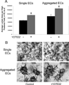
Figure 11. Rho kinase inhibitors enhance EC lumen formation of single and pre-aggregated ECs in 3D collagen matrices
ECs were either suspended within 3D collagen matrices as single cells or pre-aggregated cells in the presence or absence of 10 mM Y27632 to block Rho kinase activity. Cultures were fixed at 24 hr and were either photographed (B) or quantitated for lumen and tube area (A). Arrowheads indicate EC lumen structures. p< 0.01, n> 10 fields from triplicate cultures.
Figure 12
Figure 12. Blockade of JamB and JamC molecules using neutralizing antibodies markedly inhibits lumen and tube formation of endothelial cells
ECs were seeded within collagen type I matrices in the presence or absence of blocking antibodies to JamB, JamC either alone or blocking antibodies to α5 or α2 integrin subunits. Each antibody was added at 50µg/ml and the cultures were fixed, stained and photographed after 24 hr. Arrowheads indicate EC lumenal structures, arrows indicate ECs without lumens. Bar equals 50 µm.
Figure 13
Figure 13. MT1-MMP activity is required for EC lumen and tube formation and generation of vascular guidance tunnels in 3D collagen matrices
(A) Representative images from 3D cultures of ECs seeded within collagen type I matrices in the presence or absence of recombinant TIMP-1 or TIMP-3 added at 5 µg/ml. Cultures were fixed, stained and photographed at 24 hr. White arrowheads indicate EC lumenal structures. Bar equals 50 µm. (B) Representative images from 3D cultures of ECs seeded within collagen type I matrices after having undergone treatment with the indicated siRNAs. Cultures were fixed, stained and photographed at 24 hr. White arrowheads indicate EC lumenal structures. Bar equals 50 µm. (C) Representative images of ECs seeded within FITC-labeled collagen type I matrices in the presence or absence of the proteinase inhibitor GM6001 added at 5 µM. Cultures were fixed and photographed using confocal microscopy at 24hrs. White arrowheads indicate vascular guidance tunnels generated as a consequence of the EC lumen and tube formation. Bar equals 100 µm.
Figure 14
Figure 14. Mechanisms controlling of EC-pericyte tube stabilization in 3D extracellular matrices
A schematic diagram is shown that describes factors and mechanisms that control vascular tube stabilization in response to EC-pericyte interactions. Pericyte recruitment to tubes leads to vascular basement membrane matrix assembly which occurs within EC-generated vascular guidance tunnels. The pericyte-derived factors, TIMP-3 and angiopoietin-1 and the EC-derived factors, TIMP-2, PDGF-BB and HB-EGF, play a role in both pericyte recruitment and stabilization events. TIMP-2 and TIMP-3 together block both EC tube formation as well as regression events and also promote stabilization by inhibition of VEGFR2.

References

    1. Adams RH, Alitalo K. Molecular regulation of angiogenesis and lymphangiogenesis. Nat Rev Mol Cell Biol. 2007;8:464–478. - PubMed
    1. Alajati A, Laib AM, Weber H, Boos AM, Bartol A, Ikenberg K, Korff T, Zentgraf H, Obodozie C, Graeser R, Christian S, Finkenzeller G, Stark GB, Heroult M, Augustin HG. Spheroid-based engineering of a human vasculature in mice. Nat Methods. 2008;5:439–445. - PubMed
    1. Alavi A, Hood JD, Frausto R, Stupack DG, Cheresh DA. Role of Raf in vascular protection from distinct apoptotic stimuli. Science. 2003;301:94–96. - PubMed
    1. Aplin AC, Fogel E, Zorzi P, Nicosia RF. The aortic ring model of angiogenesis. Methods Enzymol. 2008;443:119–136. - PubMed
    1. Apte SS. A disintegrin-like and metalloprotease (reprolysin-type) with thrombospondin type 1 motif (ADAMTS) superfamily: functions and mechanisms. J Biol Chem. 2009;284:31493–31497. - PMC - PubMed

MeSH terms

Substances