. 2011 Aug;39(14):6186-200.
doi: 10.1093/nar/gkr189. Epub 2011 Apr 10.

Activity of the human immunodeficiency virus type 1 cell cycle-dependent internal ribosomal entry site is modulated by IRES trans-acting factors

Affiliations

Activity of the human immunodeficiency virus type 1 cell cycle-dependent internal ribosomal entry site is modulated by IRES trans-acting factors

Maricarmen Vallejos et al. Nucleic Acids Res. 2011 Aug.

Abstract

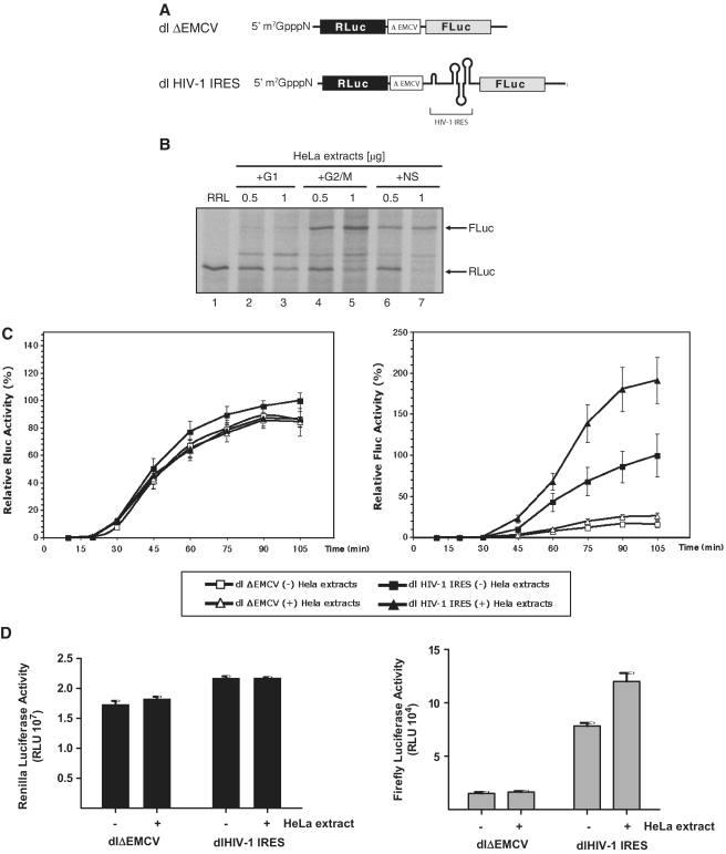
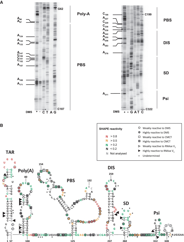
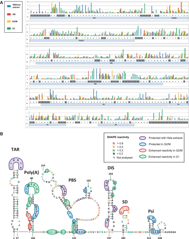
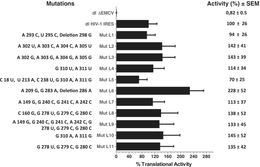
The 5' leader of the human immunodeficiency virus type 1 (HIV-1) genomic RNA harbors an internal ribosome entry site (IRES) that is functional during the G2/M phase of the cell cycle. Here we show that translation initiation mediated by the HIV-1 IRES requires the participation of trans-acting cellular factors other than the canonical translational machinery. We used 'standard' chemical and enzymatic probes and an 'RNA SHAPE' analysis to model the structure of the HIV-1 5' leader and we show, by means of a footprinting assay, that G2/M extracts provide protections to regions previously identified as crucial for HIV-1 IRES activity. We also assessed the impact of mutations on IRES function. Strikingly, mutations did not significantly affect IRES activity suggesting that the requirement for pre-formed stable secondary or tertiary structure within the HIV-1 IRES may not be as strict as has been described for other viral IRESes. Finally, we used a proteomic approach to identify cellular proteins within the G2/M extracts that interact with the HIV-1 5' leader. Together, data show that HIV-1 IRES-mediated translation initiation is modulated by cellular proteins.


Figures

Figure 1.
Figure 1.
HeLa cell cytoplasmic factors are required for HIV-1 IRES activity. (A) Schematic representation of the dl HIV-1 IRES and dlΔEMCV RNAs used in this study. (B) RRL alone or supplemented with cytoplasmic extracts (0.5 or 1 µg of total protein) generated from NS HeLa cells or cells arrested in G1 or in G2/M were programmed with the dl HIV-1 IRES RNA. [35S]-methionine-labeled proteins were resolved by SDS–PAGE and visualized as indicated in ‘Materials and Methods’ section. (C) Kinetics of HIV-1 IRES translation in the presence of G2/M HeLa extracts. The capped dlΔEMCV (open shapes) or dl HIV-1 IRES (filled shapes) RNAs (8 ng/µl) were used to program RRL. In vitro translation reactions were supplemented with 160 ng/µl of G2/M cytoplasmic extracts (Δ). Renilla luciferase (RLuc) and Firefly luciferase (FLuc) activities were measured at the indicated times. The RLuc and FLuc activities of the dl HIV-1 IRES measured at 105 min of in vitro translation in non-suplemented RRL (filled square) were arbitrary set to 100%. Relative RLuc activity (left panel) and relative FLuc activity (right panel) are shown. Values are the means ± SEM (error bars) of five independent experiments. (D) Capped and polyadenylated RNA corresponding to the dlΔEMCV or dl HIV-1 IRES vectors (6.25 ng) were microinjected into X. laevis oocytes with (+) or without (−) cytoplasmic extracts generated from G2/M arrested HeLa cells (200 ng) as described in ‘Materials and Methods’ section. Oocytes were harvested 24 h after the microinjection and processed and RLuc and FLuc activities were determined RLU. The RLuc (left panel) and FLuc (right panel) activities for each RNA are shown. Each value is the mean ± SEM from at least three oocytes obtained from different animals.
Figure 2.
Figure 2.
Secondary structure model of the HIV-1 leader. The HIV-1 5′ leader recovered from the dl HIV IRES construct (nucleotide 1–336 from clone pNL4.3 followed by 58 nts of fLuc gene) was probed using DiMethyl Sulfate (DMS), N-Cyclohexyl-N- [N-Methylmorpholino)-ethyl]-Carbodiimide-4-Toluolsulfonate (CMCT) and RNAse V1 as previously described (12,32) or using 1-methyl-7-nitroisatoic anhydride (1M7) as a modifying agent. (A) Typical examples of probing DMS probing. The HIV-1 5′ leader was probed using (+) DMS. Reverse transcription (RT) products were separated on a 8% gel as indicated in the ‘Materials and Methods’ section. Sequencing lanes were also included. Note that DMS induces a premature RT stop 1 nt before the hit. Therefore the DMS induced stops migrate faster than the corresponding sequence product (12,32). The RT pattern of the modified RNA was compared to the profile obtained with an unmodified RNA. Some hits are indicated in the figure. The asterisks on the gel denote the nucleotide position. (B) Results were fitted in a model of the HIV-1 5′ leader (43), the respective reactivity of the different probes is indicated as motioned in the box. The main HIV-1 structural elements present in the 5′ leader the TAR and poly(A) loops, PBS, DIS, SD and Psi are indicated (43).
Figure 3.
Figure 3.
HeLa cell extracts alter the accessibility of 1M7 to single-stranded regions present within the HIV-1 5′ leader. The HIV-1 5′ leader RNA was probed using 1M7 in the presence or absence of HeLa cell cytoplasmic extracts generated from NS, or cells arrested in the G1, or in the G2/M phase of the cell cycle. (A) Histogram representing the ‘SHAPE’ reactivity for each nucleotides of the 5′-UTR in absence of extracts [(-) blue bars] in presence of NS (red bars), G1 arrested [(G1) green bars] or G2/M arrested [(G2M) yellow bars] HeLa extracts. Nucleotides which reactivity is undetermined in at least one of the tested conditions are boxed in grey. Local RNA structures are indicated as hallmark. (B) Data presented in A, are incorporated in the model depicted in Figure 2, the nucleotides protected by NS, G1 and G2/M extracts are boxed in purple, those specifically protected by G2/M extracts are boxed in blue, while those which reactivity is enhanced in presence of G2/M cytoplasmic extracts are boxed in red, nucleotides which reactivity is enhanced in presence of G1 extracts are boxed in green. The reactivity of each nucleotide in presence of G2/M extract is encoded by a specific colour, the reactivity are thus also valid for the area protected by all extracts (boxed in purple), but not for the nucleotides which reactivity is enhanced in presence of G1 extracts.
Figure 4.
Figure 4.
Translational activity of the mutant HIV-1 IRES elements. HIV-1 mutant leaders generated as described in ‘Materials and Methods’ section and arbitrarily named Mut L1–L11 were cloned into the dual luciferase bicistronic vector (dl-vector). The dl HIV-1 mutant vectors (Mut L1–L11) were transfected into HeLa cells and IRES-mediated translational activity was evaluated in comparison with the dl HIV-1 IRES vector and the dlΔEMCV control (10,41). The nucleotide changes with respect to the pNL4.3 (AF 324493) leader included in the dl HIV-1 IRES vector, are shown (left). RLuc and FLuc activities were measured and the [(FLuc/RLuc)] ratio was used as an index of IRES activity. The [(FLuc/RLuc)] ratio of the dl HIV-1 IRES vector (10), was arbitrarily set to 100%. Values are the means ±SEM (error bars) of three independent experiments.

References

    1. Jackson RJ, Hellen CU, Pestova TV. The mechanism of eukaryotic translation initiation and principles of its regulation. Nat. Rev. Mol. Cell Biol. 2010;11:113–127. - PMC - PubMed
    1. Sonenberg N, Hinnebusch AG. Regulation of translation initiation in eukaryotes: mechanisms and biological targets. Cell. 2009;136:731–745. - PMC - PubMed
    1. Lopez-Lastra M, Rivas A, Barria MI. Protein synthesis in eukaryotes: the growing biological relevance of cap-independent translation initiation. Biol. Res. 2005;38:121–146. - PubMed
    1. Fitzgerald KD, Semler BL. Bridging IRES elements in mRNAs to the eukaryotic translation apparatus. Biochim. Biophys. Acta. 2009;1789:518–528. - PMC - PubMed
    1. Jang SK, Krausslich HG, Nicklin MJ, Duke GM, Palmenberg AC, Wimmer E. A segment of the 5′ nontranslated region of encephalomyocarditis virus RNA directs internal entry of ribosomes during in vitro translation. J. Virol. 1988;62:2636–2643. - PMC - PubMed

Publication types