. 2011 Jun;134(Pt 6):1591-609.
doi: 10.1093/brain/awr039. Epub 2011 Apr 10.

Harnessing neuroplasticity for clinical applications

Affiliations

Harnessing neuroplasticity for clinical applications

Steven C Cramer et al. Brain. 2011 Jun.

Abstract

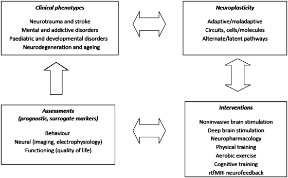
Neuroplasticity can be defined as the ability of the nervous system to respond to intrinsic or extrinsic stimuli by reorganizing its structure, function and connections. Major advances in the understanding of neuroplasticity have to date yielded few established interventions. To advance the translation of neuroplasticity research towards clinical applications, the National Institutes of Health Blueprint for Neuroscience Research sponsored a workshop in 2009. Basic and clinical researchers in disciplines from central nervous system injury/stroke, mental/addictive disorders, paediatric/developmental disorders and neurodegeneration/ageing identified cardinal examples of neuroplasticity, underlying mechanisms, therapeutic implications and common denominators. Promising therapies that may enhance training-induced cognitive and motor learning, such as brain stimulation and neuropharmacological interventions, were identified, along with questions of how best to use this body of information to reduce human disability. Improved understanding of adaptive mechanisms at every level, from molecules to synapses, to networks, to behaviour, can be gained from iterative collaborations between basic and clinical researchers. Lessons can be gleaned from studying fields related to plasticity, such as development, critical periods, learning and response to disease. Improved means of assessing neuroplasticity in humans, including biomarkers for predicting and monitoring treatment response, are needed. Neuroplasticity occurs with many variations, in many forms, and in many contexts. However, common themes in plasticity that emerge across diverse central nervous system conditions include experience dependence, time sensitivity and the importance of motivation and attention. Integration of information across disciplines should enhance opportunities for the translation of neuroplasticity and circuit retraining research into effective clinical therapies.


Figures

Figure 1
Figure 1
Conceptual overview of the relationship between clinical phenotypes, neuroplasticity, therapeutic interventions and assessment of function.
Figure 2
Figure 2
Studies of the upper extremity motor system after stroke illustrate a number of forms of brain plasticity. (A–D) Brain plasticity associated with spontaneous recovery. Several patterns of change arise spontaneously during the weeks following stroke onset. Laterality is reduced. Normally, unilateral arm movement is associated with activity mainly in the contralateral hemisphere, but after a stroke, activity is often seen in both hemispheres. Activity also increases in multiple brain areas throughout the motor network. These two points are demonstrated in A by Fujii and Nakada, (2003), who used functional MRI and found that right hand grasping movements produced predominantly left motor cortex activation in a healthy control; but in a typical patient with right hemiparesis, these movements were associated with a shift in motor cortex laterality towards the right hemisphere (double arrow) as well as increased recruitment of left dorsal premotor cortex (single arrow) and bilateral supplementary motor area (arrowhead). These patterns occur along a gradient. Thus, in general, the poorer the behavioural outcome, the more these two mechanisms are invoked. For example, (B) shows data from Ward et al. (2003). Across 20 patients, those with poorer recovery were more likely to recruit a number of bilateral motor-related brain regions over and above those seen in the control group during a functional MRI grip task by the paretic hand, whereas patients with more complete recovery were more likely to have a normal pattern of brain activation. Consistent with this (C), a poorer behavioural outcome is associated with a smaller volume of excitable motor cortex. Thickbroom et al. (2004), who used transcranial magnetic stimulation, found a linear relationship between the paretic hand's grip strength and the size of its motor cortex map, each measure expressed as a ratio of values from the contralesional side. Another pattern of spontaneous post-stroke plasticity is a shift in the location of primary sensorimotor cortex activity. A number of patterns of map reorganization have been described after stroke; for the upper extremity motor representational map, a posterior or a ventral shift has been described most often. In (D), Weiller et al. (1993), using a PET measure of regional cerebral blood flow, demonstrated that in patients with capsular infarct such as the case depicted, motor cortex activity extended from the hand area (arrowhead) ventrally into the face area (arrow). (E–G) Brain plasticity associated with treatment-induced recovery. A number of training paradigms have been introduced to patients in the chronic, plateau phase of stroke. Behavioural gains in the affected arm in this context are in general associated with an increase in the volume of activity and excitability of motor cortex, as well as an increase in laterality, back towards normal, i.e. with a greater predominance of activity in stroke-affected motor cortex rather than bilateral activation. In (E), Takahashi et al. (2008), using serial functional MRI scans across 3 weeks of robot-based physiotherapy in patients with stroke affecting the left brain, found that therapy centred around right (paretic) hand grasping movements was associated with a >20-fold increase in left (stroke-affected) sensorimotor cortex (arrow); some specificity of treatment effect was apparent given absence of significant change in the map for supination, a similar but non-rehearsed task. Similar results have been described across treatment modalities. (F) Tardy et al. (2006) found that the stimulant methylphenidate improved motor performance in the paretic hand, which was paralleled by increased functional MRI activation in sites that included stroke-affected primary sensorimotor cortex (arrow). Laterality also changes with treatment. (G) Carey et al. (2002), using functional MRI, found that training of the paretic finger was associated with an increase in the primary sensorimotor cortex laterality index during performance of the trained movement. The laterality index varies from +1 (all sensorimotor cortex activation is contralateral to movement) to –1 (all activation is ipsilateral to movement). Prior to therapy, the value for stroke patients was –0.26. After therapy, the value increased to +0.43, reflecting a shift of activation towards the stroke-affected hemisphere during paretic finger movement, and matching more closely the values observed in treated healthy control subjects.

Comment in

References

    1. Awaya S. Stimulus deprivation amblyopia in humans. In: Reinecke R, editor. Strabismus. New York: Grune and Stratton; 1987.
    1. Aylward EH, Richards TL, Berninger VW, Nagy WE, Field KM, Grimme AC, et al. Instructional treatment associated with changes in brain activation in children with dyslexia. Neurology. 2003;61:212–9. - PubMed
    1. Bagati D, Nizamie SH, Prakash R. Effect of augmentatory repetitive transcranial magnetic stimulation on auditory hallucinations in schizophrenia: randomized controlled study. Aust N Z J Psychiatry. 2009;43:386–92. - PubMed
    1. Baillieux H, De Smet HJ, Dobbeleir A, Paquier PF, De Deyn PP, Marien P. Cognitive and affective disturbances following focal cerebellar damage in adults: a neuropsychological and SPECT study. Cortex. 2010;46:869–79. - PubMed
    1. Balu DT, Coyle JT. Neuroplasticity signaling pathways linked to the pathophysiology of schizophrenia. Neurosci Biobehav Rev. 2011;35:848–70. - PMC - PubMed

Publication types