. 2011 Jul;25(7):2456-67.
doi: 10.1096/fj.10-176669. Epub 2011 Apr 11.

Cardiac ATP-sensitive K+ channel associates with the glycolytic enzyme complex

Affiliations

Cardiac ATP-sensitive K+ channel associates with the glycolytic enzyme complex

Miyoun Hong et al. FASEB J. 2011 Jul.

Abstract

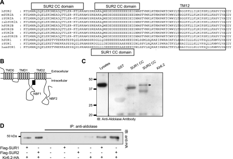
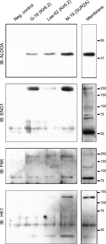
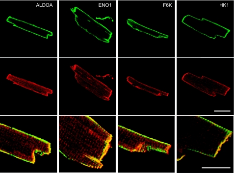
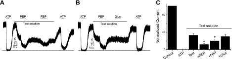
Being gated by high-energy nucleotides, cardiac ATP-sensitive potassium (K(ATP)) channels are exquisitely sensitive to changes in cellular energy metabolism. An emerging view is that proteins associated with the K(ATP) channel provide an additional layer of regulation. Using putative sulfonylurea receptor (SUR) coiled-coil domains as baits in a 2-hybrid screen against a rat cardiac cDNA library, we identified glycolytic enzymes (GAPDH and aldolase A) as putative interacting proteins. Interaction between aldolase and SUR was confirmed using GST pulldown assays and coimmunoprecipitation assays. Mass spectrometry of proteins from K(ATP) channel immunoprecipitates of rat cardiac membranes identified glycolysis as the most enriched biological process. Coimmunoprecipitation assays confirmed interaction for several glycolytic enzymes throughout the glycolytic pathway. Immunocytochemistry colocalized many of these enzymes with K(ATP) channel subunits in rat cardiac myocytes. The catalytic activities of aldolase and pyruvate kinase functionally modulate K(ATP) channels in patch-clamp experiments, whereas D-glucose was without effect. Overall, our data demonstrate close physical association and functional interaction of the glycolytic process (particularly the distal ATP-generating steps) with cardiac K(ATP) channels.


Figures

Figure 1.
Figure 1.
Identification of putative SUR CC domains and interaction with aldolase. A) Using the COILS computer algorithm, we identified several putative CC domains in both SUR1 and SUR2 subunits. See Supplemental Fig. S1 and Supplemental Table S1 for more information. Sequences are shown for human SUR2A (hSUR2), mouse SUR2A (mSUR2A), rabbit SUR2A (rabSUR2A), human SUR2B (hSUR2B), mouse SUR2B (mSUR2B), rabbit SUR2B (rabSUR2B), human SUR1 (hSUR1), rat SUR1 (rSUR1), and hamster SUR1 (hamSUR1). B) SUR subunit topology, depicting the location of the putative CC domains (black box). C) Aldolase interacts with the SUR CC domains. A GST pulldown experiment was performed using GST alone, GST-SUR1 CC domain, GST-SUR2 CC domain, and the GST-Kir6.2 C terminus. Immunoblotting was performed using an anti-aldolase antibody. Left lane: cell lysates (no pulldown). D) COS7 cells were transfected with cDNA combinations as shown. Each protein was expressed in cell lysates (not shown). Immunoprecipitates obtained with an anti-aldolase antibody (C-16) were separated by SDS-PAGE and immunoblotted with an anti-HA antibody.
Figure 2.
Figure 2.
Coimmunoprecipitation of glycolytic enzymes and KATP channel subunits in rat heart membranes. KATP channel subunits were immunoprecipitated using anti-Kir6.2 antibodies (G-16 or Lee-62) or antibodies directed against the SUR2A subunit (M-19) or an unrelated IgG as a negative control (see Materials and Methods for full details). Immunoprecipitates were subjected to SDS-PAGE and immunoblotted with monoclonal antibodies against the glycolytic enzymes ALDOA, ENO1, F6K, or HK1. Right lanes: depiction of the presence of the proteins in membranes (no immunoprecipitation).
Figure 3.
Figure 3.
Coimmunostaining of glycolytic enzymes and Kir6.2 in rat cardiomyocytes. Top panels: localization of ALDOA, ENO1, F6K, and HK1 using monoclonal antibodies. Detection was in the green channel (DyLight 488). Middle panels: staining with the C-62 anti-Kir6.2 antibody (detected in the red channel; DyLight 549). Bottom panels: overlays of the red and green channels. Overlay image is a magnified region of the top 2 images to show more detail. Scale bars = 40 μm (top and middle panels); 20 μm (bottom panels).
Figure 4.
Figure 4.
Substrates of PK and ALDOA, but not glucose, suppress KATP channel in excised, inside-out membrane patches. A) Channel activity was blocked by ATP (1 mM) and was partially inhibited by the test solution (containing 90 μM ATP, 0.5 mM ADP, 1 mM KH2PO4, and 1 mM NAD+). Further addition of PEP (5 mM) and FBP (5 mM) reversibly inhibited channel activity. B) Similar experiment depicting the effects of PEP and glucose (Gluc; 10 mM). C) Summary data (means±se) normalized to control current (in the absence of ATP). Dotted lines indicate zero current.

References

    1. Ashcroft F. M. (1988) Adenosine 5′-triphosphate-sensitive potassium channels. Annu. Rev. Neurosci. 11, 97–118 - PubMed
    1. Nichols C. G. (2006) KATP channels as molecular sensors of cellular metabolism. Nature 440, 470–476 - PubMed
    1. Weiss J. N., Lamp S. T. (1989) Cardiac ATP-sensitive K + channels - evidence for preferential regulation by glycolysis. J. Gen. Physiol. 94, 911–935 - PMC - PubMed
    1. Dubinsky W. P., Mayorga-Wark O., Schultz S. G. (1998) Colocalization of glycolytic enzyme activity and KATP channels in basolateral membrane of Necturus enterocytes. Am. J. Physiol. 275, C1653–C1659 - PubMed
    1. Dhar Chowdhury P., Harrell M. D., Han S., Jankowska D., Parachuru L., Morrissey A., Srivastava S., Liu W., Malester B., Yoshida H., Coetzee W. A. (2005) The glycolytic enzymes, glyceraldehyde-3-phosphate dehydrogenase, triose phosphate isomerase and pyruvate kinase are components of the KATP channel macromolecular complex and regulate its function. J. Biol. Chem. 280, 38464–38470 - PMC - PubMed

Publication types

MeSH terms

Substances

LinkOut - more resources