. 2011 Jun;31(11):2210-26.
doi: 10.1128/MCB.00964-10. Epub 2011 Apr 11.

IkappaB kinase beta promotes cell survival by antagonizing p53 functions through DeltaNp73alpha phosphorylation and stabilization

Affiliations

IkappaB kinase beta promotes cell survival by antagonizing p53 functions through DeltaNp73alpha phosphorylation and stabilization

Rosita Accardi et al. Mol Cell Biol. 2011 Jun.

Abstract

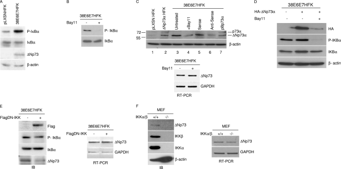
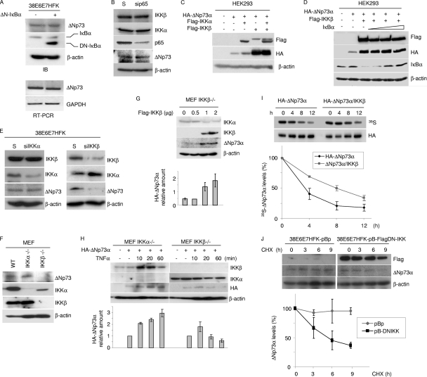
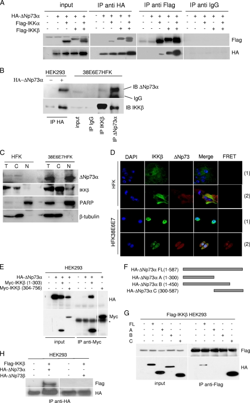
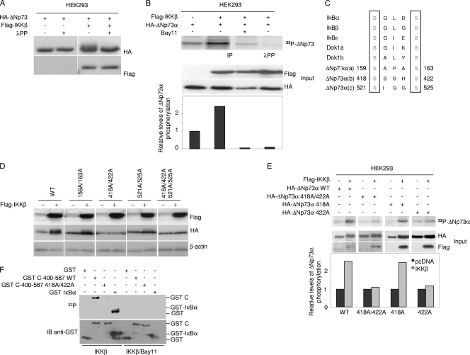
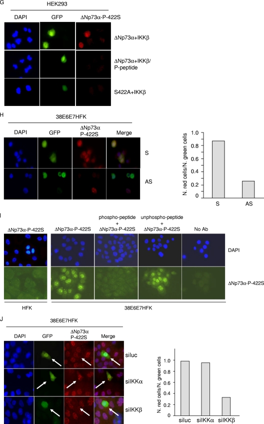
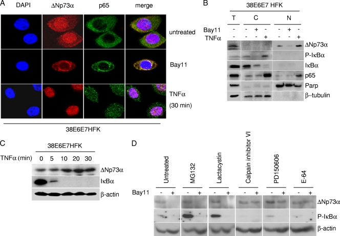
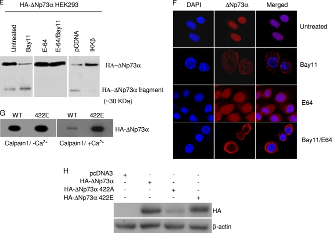
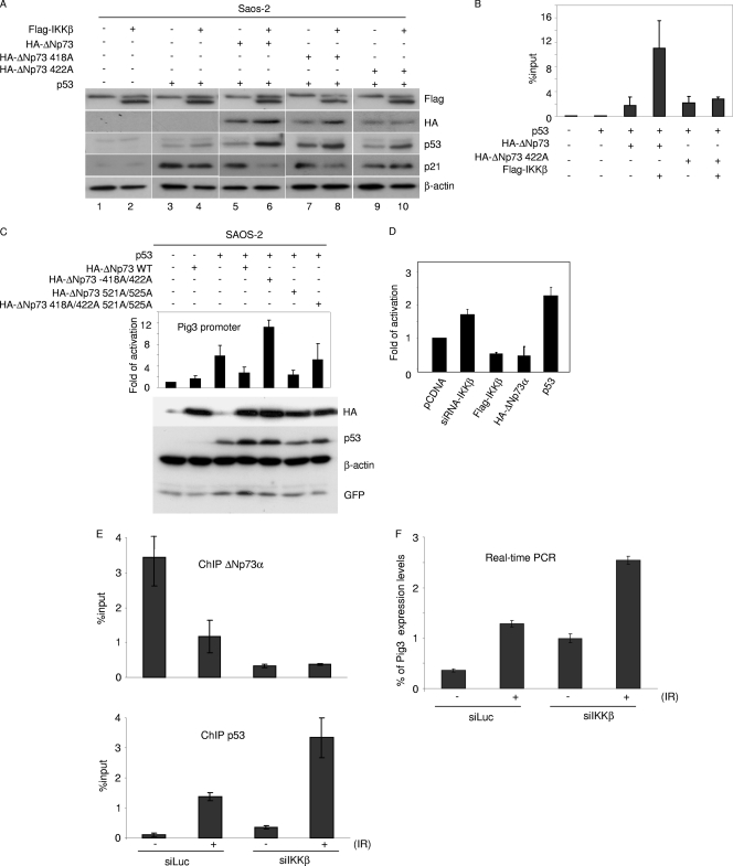
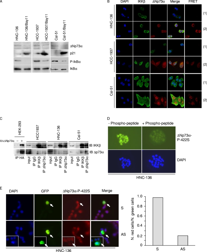
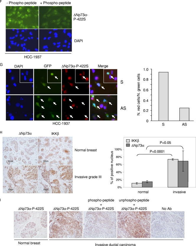
ΔNp73α, a dominant-negative inhibitor of p53 and p73, exhibits antiapoptotic and transforming activity in in vitro models and is often found to be upregulated in human cancers. The mechanisms involved in the regulation of ΔNp73α protein levels in normal and cancer cells are poorly characterized. Here, we show that that IκB kinase beta (IKKβ) increases ΔNp73α protein stability independently of its ability to activate NF-κB. IKKβ associates with and phosphorylates ΔNp73α at serine 422 (S422), leading to its accumulation in the nucleus, where it binds and represses several p53-regulated genes. S422A mutation in ΔNp73α abolished IKKβ-mediated stabilization and inhibition of p53-regulated gene expression. Inhibition of IKKβ activity by chemical inhibitors, overexpression of dominant-negative mutants, or gene silencing by siRNA also resulted in ΔNp73α destabilization, which under these conditions was rapidly translocated into the cytoplasm and degraded by a calpain-mediated mechanism. We also present evidence for the IKKβ and ΔNp73α cross talk in cancer-derived cell lines and primary cancers. Our data unveil a new mechanism involved in the regulation of the p73 and p53 network.


Figures

Fig. 1.
Fig. 1.
The IKK complex regulates the stability of the ΔNp73 protein. (A) Protein extracts of primary keratinocytes transduced with empty retrovirus vector (pLXSN HFK) or immortalized keratinocytes expressing HPV type 38 E6 and E7 (38E6E7HFK) were analyzed by immunoblotting with the indicated antibodies. (B) 38E6E7HFK cells were treated with Bay11 or dimethyl sulfoxide (DMSO) for 2 h. Protein extracts were prepared and analyzed by immunoblotting with the indicated antibodies. (C) Protein extracts of pLXSN HFKs or HFKs stably expressing ΔNp73α or p73α, 38E6E7HFK cells, or 38E6E7HFK cells cultured in the presence of Bay11 or transfected with ΔNp73 sense or antisense oligonucleotide were analyzed by immunoblotting with the indicated antibodies (upper panel). Total RNA was also extracted from Bay11-treated 38E6E7HFK cells, and ΔNp73 or GAPDH mRNA levels were measured by RT-PCR (bottom panel). (D) 38E6E7HFK cells were transfected with a HA-ΔNp73α construct. Cells were treated with Bay11 or DMSO for 2 h and processed as for panel A. (E) 38E6E7HFK cells were transduced with pBabe-puro (pBp) or pBp-flagged dominant-negative kinase-dead IKK (Flag-DN-IKK) retrovirus. Protein extracts were analyzed by immunoblotting with the indicated antibodies (left panel). Total RNA was also extracted from both cell lines, and ΔNp73 or GAPDH mRNA levels were measured by RT-PCR (right panel). (F) Protein extracts from wild-type MEFs (IKKα/β+/+) and IKKα/β−/− MEFs were prepared and analyzed by immunoblotting using the indicated antibodies (left panel). Total RNA was prepared from wild-type or IKKα/β knockout MEFs, and the levels of ΔNp73 transcript were determined by RT-PCR (right panel).
Fig. 2.
Fig. 2.
IKKβ increases ΔNp73 protein levels. (A) 38E6E7HFK cells were transduced with pBp or with pBp-ΔN-IκBα superrepressor (ΔN-IκBα). Protein extracts were analyzed by immunoblotting using the indicated antibodies (top panel). Total RNA was also extracted from both cell lines, and ΔNp73 or GAPDH mRNA levels were measured by RT-PCR (bottom panel). (B) Scrambled (S) RNA and siRNA for p65 (sip65) was transfected in 38E6E7HFK cells. Thirty-six hours after transfection, protein extracts were analyzed by immunoblotting with the indicated antibodies. (C and D) HEK293 cells were transfected with different expression constructs as indicated. After 24 h, protein extracts were analyzed by immunoblotting with the indicated antibodies. (E) Scrambled (S) RNA and siRNA for IKKα (siIKKα) or IKKβ (siIKKβ) was transfected in 38E6E7HFK cells. Thirty-six hours after transfection, protein extracts were analyzed by immunoblotting with the indicated antibodies. (F) IKKα/β+/+ (WT), IKKα−/−, and IKKβ−/− MEF cellular protein extracts were analyzed by immunoblotting with the indicated antibodies. (G) IKKβ−/− MEF cells were transfected with increasing concentrations of pcDNA3-Flag-IKKβ, and 24 h after transfection, protein extracts were analyzed by immunoblotting with the indicated antibodies (top panel). The ΔNp73 protein signal was quantified by the Quantity One software program (Bio-Rad), normalized on the levels of β-actin, and the values obtained were reported in the histogram (bottom panel). The data are the means of results from two independent experiments. (H) IKKα−/− MEF and IKKβ−/− MEF cells were transfected with pcDNA3-HA-ΔNp73α and treated with TNF-α at the indicated time points. Protein extracts were analyzed by immunoblotting with the indicated antibodies (top panel). The amounts of HA-ΔNp73 signal in the Western blot were quantified as explained for panel G and are reported in the histogram (bottom panel). The data are the means of results from two independent experiments. (I) HEK293 cells were transfected with HA-tagged ΔNp73α in the absence or presence of overexpressed Flag-tagged IKKβ. Twenty-four hours after transfection, cells were labeled for 1 h with l- [35S]methionine, chased for the indicated times, and collected. Following anti-HA immunoprecipitation, the immunocomplexes were loaded on an SDS-PAGE gel and transferred onto a polyvinylidene difluoride (PVDF) membrane. Autoradiography (top panel) and then anti-HA Western blotting (middle panel) were performed; 35S-HA-ΔNp73 bands were quantified by Image Lab (Bio-Rad) and normalized on the total levels of immunoprecipitated HA-ΔNp73 protein. The percentage of ΔNp73 at time zero was referred to as 100%, and the percentages of protein at the different time points were calculated relative to that at time zero and reported in the histogram (lower panel). The data are the means of results for two independent experiments. (J) 38E6E7HFK cells stably expressing a dominant-negative inhibitor of IKKβ (pB-FlagDN-IKK) were generated and cultured in the presence of CHX for the indicated number of hours. At each time point, cells were collected. Protein extracts were prepared and analyzed by immunoblotting with the indicated antibodies (upper panel). The levels of ΔNp73 were quantified by Quantity One (Bio-Rad) and normalized on the levels of β-actin. The percentages of ΔNp73 at the different time points were calculated as for Fig. 1G and are reported in the histogram (lower panel). The data are the means of results for two independent experiments.
Fig. 3.
Fig. 3.
IKKβ directly interacts with ΔNp73. (A) HEK293 cells were transfected with the indicated expression plasmids, and 24 h after transfection, protein extracts were subjected to immunoprecipitation followed by immunoblotting. “Input” represents 1/10 of total extracts used for the immunoprecipitation. (B) One-milligram protein extracts of 38E6E7HFK cells were immunoprecipitated with the indicated antibodies, followed by immunoblotting. “Input” represents 1/10 of total extracts used for the immunoprecipitation. As a control, immunocomplexes obtained from HEK293 cells transfected with pcDNA or pcDNA3-HA-ΔNp73α were included in the experiment. (C) Primary HFK and 38E6E7HFK cells were collected and fractionated by using a nuclear extraction kit (Panomics). After fractionation, total extract (T), cytoplasm (C), and nucleus (N) were analyzed by immunoblotting with the indicated antibodies. (D) Primary HFK cells transduced with empty retrovirus (pLXSN) or 38E6E7HFK cells were seeded on coverslips. After immunofluorescent staining for ΔNp73 and IKKβ with specific antibodies, fluorescent signal was visualized using confocal microscopy. FRET analysis was performed as explained in Materials and Methods. A representative image of a FRET positive signal for each cell line is shown in the right panels (2). As a negative control, FRET analyses were performed with cells stained only with the fluorochrome donor (Alexa 488) (1). (E) HEK293 cells were transfected with the indicated expression plasmids; 24 h posttransfection, protein extracts were immunoprecipitated and analyzed by immunoblotting. The last lane was obtained in the same experiment and was taken from a different area of the same SDS-PAGE gel. The asterisk indicates the IgG heavy chain. (F) Schematic representation of ΔNp73 deletion mutants. (G) HEK293 cells were transfected with pcDNA3 constructs expressing wild-type or deleted HA-ΔNp73 mutants together with pcDNA3-Flag-IKKβ as indicated. After 24 h, cells were collected and protein extracts were prepared. Immunoprecipitations were performed using an anti-Flag antibody, and pellets were analyzed by immunoblotting. Input represents 1/10 of total extracts used for the immunoprecipitation. (H) HEK293 cells were transfected with pcDNA3 constructs as indicated. After 24 h, protein extracts were immunoprecipitated with an HA-tag antibody, and pellets were analyzed by immunoblotting.
Fig. 4.
Fig. 4.
IKKβ phosphorylates ΔNp73. (A) HEK293 cells were transfected with expression plasmids as indicated. After 24 h, protein extracts were treated or not with λPP and analyzed by immunoblotting. (B) HEK293 cells were transfected with the indicated pcDNA3 constructs and cultured in the presence of [32P]orthophosphate with or without Bay11. Immunoprecipitations were performed using anti-HA-tag antibody. One immunopellet was treated with λ-phosphatase (top panel, fourth lane). Proteins were analyzed by autoradiography and by immunoblotting (top and lower panels, respectively). The histogram (bottom panel) shows the phosphorylation levels of the different ΔNp73α protein bands. For each band, radioactive signal was normalized to the immunoblotting signal of the input. (C) Amino acid sequence alignments of IKK phosphorylation sites of two cellular proteins that are targeted by IKK complex (IκB and Dok1) with potential IKK phosphorylation sites of ΔNp73α. Numbers indicate the positions of serines in ΔNp73α that are potentially phosphorylated by IKK. (D) HEK293 cells were transfected with wild-type ΔNp73α (WT) or various alanine substitution mutants with or without Flag-IKKβ. Protein extracts were analyzed by immunoblotting with the indicated antibodies. (E) HEK293 cells were transfected with the indicated constructs, and 32P labeling was performed as described in the legend for panel B. Immunoprecipitation was performed using anti-HA-tag antibody, followed by immunoblotting or autoradiography (top panels). The histogram (bottom panel) was obtained as explained in the legend for panel B. (F) Kinetic reactions were carried out as described in Materials and Methods using GST fusion proteins with the C-terminal domain of wild-type or 422A mutant ΔNp73α or IκBα. Radioactive and cold proteins were detected by autoradiography and immunoblotting, respectively (top and lower panels). (G) HEK293 cells were cotransfected with IKKβ and wild-type ΔNp73α or the 442A ΔNp73α mutant together with pE-GFPCI. Cells were seeded on coverslips and subjected to immunofluorescence analysis as described in Materials and Methods with the indicated antibodies. (H) 38E6E7HFK cells were transfected with S or AS against ΔNp73α together with the pE-GFPCI construct. Thirty hours posttransfection, cells were stained with anti-ΔNp73α P-422S antibody and analyzed by immunofluorescence. Representative images are shown in the left panel. Quantification of ΔNp73α P-422S and GFP-positive cells or GFP-positive cells was determined by counting at least 100 transfected cells in more than 10 different fields (right panel). (I) HFK or 38E6E7HFK cells were stained with a specific antibody against the 422S-phosphorylated form of ΔNp73α. As controls, immunofluorescence was also performed without the primary antibody (no Ab) or with primary antibody preincubated with the phosphorylated or nonphosphorylated 422S ΔNp73α peptide. (J) 38E6E7HFK cells were cotransfected with pE-GFPCI and siRNAs against IKKα (siIKKα), IKKβ (siIKKβ), or siLuc as a negative control. Thirty hours posttransfection, cells were stained with anti-ΔNp73α P-422S antibody and subjected to immunofluorescence analysis. Representative imagines are shown in the left panel; arrows indicate transfected cells. Quantification of ΔNp73α P-422S and GFP-positive cells or GFP-positive cells was determined by counting at least 100 transfected cells in more than 10 different fields (right panel).
Fig. 4.
Fig. 4.
IKKβ phosphorylates ΔNp73. (A) HEK293 cells were transfected with expression plasmids as indicated. After 24 h, protein extracts were treated or not with λPP and analyzed by immunoblotting. (B) HEK293 cells were transfected with the indicated pcDNA3 constructs and cultured in the presence of [32P]orthophosphate with or without Bay11. Immunoprecipitations were performed using anti-HA-tag antibody. One immunopellet was treated with λ-phosphatase (top panel, fourth lane). Proteins were analyzed by autoradiography and by immunoblotting (top and lower panels, respectively). The histogram (bottom panel) shows the phosphorylation levels of the different ΔNp73α protein bands. For each band, radioactive signal was normalized to the immunoblotting signal of the input. (C) Amino acid sequence alignments of IKK phosphorylation sites of two cellular proteins that are targeted by IKK complex (IκB and Dok1) with potential IKK phosphorylation sites of ΔNp73α. Numbers indicate the positions of serines in ΔNp73α that are potentially phosphorylated by IKK. (D) HEK293 cells were transfected with wild-type ΔNp73α (WT) or various alanine substitution mutants with or without Flag-IKKβ. Protein extracts were analyzed by immunoblotting with the indicated antibodies. (E) HEK293 cells were transfected with the indicated constructs, and 32P labeling was performed as described in the legend for panel B. Immunoprecipitation was performed using anti-HA-tag antibody, followed by immunoblotting or autoradiography (top panels). The histogram (bottom panel) was obtained as explained in the legend for panel B. (F) Kinetic reactions were carried out as described in Materials and Methods using GST fusion proteins with the C-terminal domain of wild-type or 422A mutant ΔNp73α or IκBα. Radioactive and cold proteins were detected by autoradiography and immunoblotting, respectively (top and lower panels). (G) HEK293 cells were cotransfected with IKKβ and wild-type ΔNp73α or the 442A ΔNp73α mutant together with pE-GFPCI. Cells were seeded on coverslips and subjected to immunofluorescence analysis as described in Materials and Methods with the indicated antibodies. (H) 38E6E7HFK cells were transfected with S or AS against ΔNp73α together with the pE-GFPCI construct. Thirty hours posttransfection, cells were stained with anti-ΔNp73α P-422S antibody and analyzed by immunofluorescence. Representative images are shown in the left panel. Quantification of ΔNp73α P-422S and GFP-positive cells or GFP-positive cells was determined by counting at least 100 transfected cells in more than 10 different fields (right panel). (I) HFK or 38E6E7HFK cells were stained with a specific antibody against the 422S-phosphorylated form of ΔNp73α. As controls, immunofluorescence was also performed without the primary antibody (no Ab) or with primary antibody preincubated with the phosphorylated or nonphosphorylated 422S ΔNp73α peptide. (J) 38E6E7HFK cells were cotransfected with pE-GFPCI and siRNAs against IKKα (siIKKα), IKKβ (siIKKβ), or siLuc as a negative control. Thirty hours posttransfection, cells were stained with anti-ΔNp73α P-422S antibody and subjected to immunofluorescence analysis. Representative imagines are shown in the left panel; arrows indicate transfected cells. Quantification of ΔNp73α P-422S and GFP-positive cells or GFP-positive cells was determined by counting at least 100 transfected cells in more than 10 different fields (right panel).
Fig. 5.
Fig. 5.
IKKβ affects stability and intracellular localization of ΔNp73α. (A) 38E6E7HFK cells were treated with TNF-α or Bay11 and ΔNp73, and p65 cellular localization was visualized by immunofluorescence with indicated antibodies. (B) 38E6E7HFK cells treated with TNF-α or Bay11 were collected and fractionated by using a nuclear extraction kit (Panomics). After fractionation, total extract (T), cytoplasm (C), and nucleus (N) were analyzed by immunoblotting with the indicated antibodies. (C) 38E6E7HFK cells were treated with TNF-α at the indicated time points. Protein extracts were prepared and analyzed by immunoblotting using the indicated antibodies. (D) 38E6E7HFK cells were treated with the indicated protease inhibitors for 8 h and Bay11 (+) or DMSO (−) for 2 h. Protein extracts were analyzed by immunoblotting. (E) HEK293 cells were transfected with the pcDNA3 HA-ΔNp73 construct and cultured under specified conditions (left and right panels) or cotransfected with different pcDNA3 constructs in the indicated combinations (right panel). Cells were then treated with indicated inhibitors followed by immunoprecipitation of whole-cell lysates and immunoblotting with the indicated antibodies. (F) 38E6E7HFK cells were seeded on coverslips and treated for 8 h with E-64 and/or for 2 h with Bay11. After the cells were fixed, ΔNp73 was visualized by immunofluorescence with specified antibodies. (G) In vitro-translated HA-tagged WT and mutant 422E ΔNp73 were incubated with recombinant calpain I in the presence or absence of Ca2+. After 30 min, the reaction was stopped by the addition of SDS loading buffer, and samples were analyzed by immunoblotting with an anti-HA-tag antibody. (H) 38E6E7HFK cells were transfected with the pcDNA3-expressing wild type or 422A or 422E HA-ΔNp73α. After 24 h, protein extracts were analyzed by immunoblotting with the indicated antibodies.
Fig. 5.
Fig. 5.
IKKβ affects stability and intracellular localization of ΔNp73α. (A) 38E6E7HFK cells were treated with TNF-α or Bay11 and ΔNp73, and p65 cellular localization was visualized by immunofluorescence with indicated antibodies. (B) 38E6E7HFK cells treated with TNF-α or Bay11 were collected and fractionated by using a nuclear extraction kit (Panomics). After fractionation, total extract (T), cytoplasm (C), and nucleus (N) were analyzed by immunoblotting with the indicated antibodies. (C) 38E6E7HFK cells were treated with TNF-α at the indicated time points. Protein extracts were prepared and analyzed by immunoblotting using the indicated antibodies. (D) 38E6E7HFK cells were treated with the indicated protease inhibitors for 8 h and Bay11 (+) or DMSO (−) for 2 h. Protein extracts were analyzed by immunoblotting. (E) HEK293 cells were transfected with the pcDNA3 HA-ΔNp73 construct and cultured under specified conditions (left and right panels) or cotransfected with different pcDNA3 constructs in the indicated combinations (right panel). Cells were then treated with indicated inhibitors followed by immunoprecipitation of whole-cell lysates and immunoblotting with the indicated antibodies. (F) 38E6E7HFK cells were seeded on coverslips and treated for 8 h with E-64 and/or for 2 h with Bay11. After the cells were fixed, ΔNp73 was visualized by immunofluorescence with specified antibodies. (G) In vitro-translated HA-tagged WT and mutant 422E ΔNp73 were incubated with recombinant calpain I in the presence or absence of Ca2+. After 30 min, the reaction was stopped by the addition of SDS loading buffer, and samples were analyzed by immunoblotting with an anti-HA-tag antibody. (H) 38E6E7HFK cells were transfected with the pcDNA3-expressing wild type or 422A or 422E HA-ΔNp73α. After 24 h, protein extracts were analyzed by immunoblotting with the indicated antibodies.
Fig. 6.
Fig. 6.
IKK-mediated phosphorylation of ΔNp73α increases its p53 inhibitory activity. (A) Saos-2 cells were transfected with different pcDNA3 constructs in the indicated combinations. After 24 h, protein extracts were analyzed by immunoblotting with the indicated antibodies. (B) Saos-2 cells were transfected with different pcDNA3 constructs in the indicated combinations. After 36 h, ChIP was performed using an anti-HA-tag antibody and followed by real-time PCR, using primers flanking the p53 RE within the p21 promoter. Simultaneously, 1/10 of the total chromatin was processed. The values in the histogram were obtained by dividing for each sample the amount of p21 promoter which is bound by ΔNp73-HA by the total amount of p21 promoter present in the input. (C) Saos-2 cells were transfected with the following constructs: Pig3prom-firefly luciferase reporter construct, a constitutively expressing Renilla construct, pcDNA3-HA-ΔNp73α (wild type or 418A/422A, 521A/525A, or 418A/422A/521A/525A mutant), pcDNA-p53, and pE-GFPCI. After 24 h, cells were collected and processed for the luciferase assay as described in Materials and Methods (upper panel). In parallel, protein extracts were prepared and analyzed by immunoblotting with the indicated antibodies (lower panel). (D) 38E6E7HFK cells were transfected with the following constructs: Pig3prom-firefly luciferase reporter construct and a constitutively expressing Renilla construct, in combination with siRNA-IKKβ, Flag-IKKβ, pCDNA HA-ΔNp73α, or pcDNA-p53. Thirty-six hours posttransfection, cells were collected and lysed. Luciferase activity was measured and expressed as fold activation in comparison to that of the control (pCDNA). The variation in fold activities between the different conditions was significant (P < 0.05). (E and F) 38E6E7HFK cells were transfected with siIKKβ or siLuc as a negative control; 24 h posttransfection, cells were treated with ionizing radiations (IR) (30 Gy). After irradiation, cells were allowed to grow for 8 h and processed for ChIP (E) or gene expression analysis (F). ChIP followed by real-time PCR was performed using an anti-ΔNp73 antibody (upper panel) or anti-p53 antibody (lower panel) and with primers flanking the p53-RE within the pig3 promoter. Simultaneously, 1/10 of the total chromatin was processed. The values reported in the histogram were obtained as for panel B (E). Pig3 mRNA levels in cells subjected to the indicated treatments were determined by real-time RT-PCR (F).
Fig. 7.
Fig. 7.
IKKβ and ΔNp73α cross talk in cancer-derived cell lines and primary cancers. (A) HNC-136, HCC-1937, and Cal-51 cell lines were treated with Bay11 for 2 h, and total protein extracts were analyzed by immunoblotting with the indicated antibodies. (B) HCC-1937, HNC-136, and Cal-51 cells were stained with the indicated antibodies. Fluorescent staining was visualized by confocal microscopy. FRET positive signals (2) and FRET negative controls (1) are shown in the right panels. (C) One-milligram protein extracts of HCC-1937, HNC-136, and Cal-51 were immunoprecipitated with an anti-IKKβ or -ΔNp73α antibody. As negative controls, the same amounts of total extracts from each cell line were immunoprecipitated with anti-IgG. Immunocomplexes were analyzed by immunoblotting. One-tenth of total extracts used for the immunoprecipitation was loaded as input. As a control, immunocomplexes obtained from HEK293 cells transfected with pcDNA or pcDNA3-HA-ΔNp73α were included in the experiment. (D) HNC-136 cells were stained with the indicated antibodies. Fluorescent staining was visualized by confocal microscopy. As a control, primary antibody was preincubated with an excess of 422S-phosphorylated peptide. (E) HNC-136 cells were cotransfected with pE-GFPCI and S or AS against ΔNp73α. Thirty hours posttransfection, cells were stained with anti-ΔNp73α P-422S antibody and analyzed for immunofluorescence. Representative images are shown in the left panel; arrows indicate transfected cells. The white frame indicates a different field. Quantification of ΔNp73α P-422S and GFP-positive cells or GFP-positive cells was determined by counting at least 100 transfected cells in more than 10 different fields (right panel). (F) HCC-1937 cells were stained as explained in the legend for panel D. (G) HCC-1937 cells were transfected and processed as explained in the legend for panel E. Representative images are shown in the left panel; arrows indicate transfected cells. The white frame indicates a different field. Quantification of ΔNp73α P-422S and GFP-positive cells or GFP-positive cells was determined by counting at least 100 transfected cells in more than 10 different fields (right panel). (H) ΔNp73 and IKKβ cellular localization was analyzed in normal and cancer breast tissues (left panel, representative staining). The histogram (right panel) shows the quantification of the percentage of cells in normal (n = 1) or cancer tissues (n = 3) with nuclear staining for IKKβ and ΔNp73α. (I) Normal and breast cancer tissues were stained with a specific antibody against the 422S-phosphorylated form of ΔNp73α. As controls, immunostaining was also performed without the primary antibody (no Ab) or with primary antibody preincubated with a phosphorylated or nonphosphorylated 422S ΔNp73α peptide.
Fig. 7.
Fig. 7.
IKKβ and ΔNp73α cross talk in cancer-derived cell lines and primary cancers. (A) HNC-136, HCC-1937, and Cal-51 cell lines were treated with Bay11 for 2 h, and total protein extracts were analyzed by immunoblotting with the indicated antibodies. (B) HCC-1937, HNC-136, and Cal-51 cells were stained with the indicated antibodies. Fluorescent staining was visualized by confocal microscopy. FRET positive signals (2) and FRET negative controls (1) are shown in the right panels. (C) One-milligram protein extracts of HCC-1937, HNC-136, and Cal-51 were immunoprecipitated with an anti-IKKβ or -ΔNp73α antibody. As negative controls, the same amounts of total extracts from each cell line were immunoprecipitated with anti-IgG. Immunocomplexes were analyzed by immunoblotting. One-tenth of total extracts used for the immunoprecipitation was loaded as input. As a control, immunocomplexes obtained from HEK293 cells transfected with pcDNA or pcDNA3-HA-ΔNp73α were included in the experiment. (D) HNC-136 cells were stained with the indicated antibodies. Fluorescent staining was visualized by confocal microscopy. As a control, primary antibody was preincubated with an excess of 422S-phosphorylated peptide. (E) HNC-136 cells were cotransfected with pE-GFPCI and S or AS against ΔNp73α. Thirty hours posttransfection, cells were stained with anti-ΔNp73α P-422S antibody and analyzed for immunofluorescence. Representative images are shown in the left panel; arrows indicate transfected cells. The white frame indicates a different field. Quantification of ΔNp73α P-422S and GFP-positive cells or GFP-positive cells was determined by counting at least 100 transfected cells in more than 10 different fields (right panel). (F) HCC-1937 cells were stained as explained in the legend for panel D. (G) HCC-1937 cells were transfected and processed as explained in the legend for panel E. Representative images are shown in the left panel; arrows indicate transfected cells. The white frame indicates a different field. Quantification of ΔNp73α P-422S and GFP-positive cells or GFP-positive cells was determined by counting at least 100 transfected cells in more than 10 different fields (right panel). (H) ΔNp73 and IKKβ cellular localization was analyzed in normal and cancer breast tissues (left panel, representative staining). The histogram (right panel) shows the quantification of the percentage of cells in normal (n = 1) or cancer tissues (n = 3) with nuclear staining for IKKβ and ΔNp73α. (I) Normal and breast cancer tissues were stained with a specific antibody against the 422S-phosphorylated form of ΔNp73α. As controls, immunostaining was also performed without the primary antibody (no Ab) or with primary antibody preincubated with a phosphorylated or nonphosphorylated 422S ΔNp73α peptide.

References

    1. Accardi R., et al. 2006. Skin human papillomavirus type 38 alters p53 functions by accumulation of deltaNp73. EMBO Rep. 7:334–340 - PMC - PubMed
    1. Agami R., Blandino G., Oren M., Shaul Y. 1999. Interaction of c-Abl and p73alpha and their collaboration to induce apoptosis. Nature 399:809–813 - PubMed
    1. Anest V., et al. 2003. A nucleosomal function for IkappaB kinase-alpha in NF-kappaB-dependent gene expression. Nature 423:659–663 - PubMed
    1. Buhlmann S., Putzer B. M. 2008. DNp73 a matter of cancer: mechanisms and clinical implications. Biochim. Biophys. Acta 1785:207–216 - PubMed
    1. Caldeira S., et al. 2003. The E6 and E7 proteins of cutaneous human papillomavirus type 38 display transforming properties. J. Virol. 77:2195–2206 - PMC - PubMed

Publication types

MeSH terms