. 2011 Jun;31(12):2413-21.
doi: 10.1128/MCB.05242-11. Epub 2011 Apr 11.

Mediator influences telomeric silencing and cellular life span

Affiliations

Mediator influences telomeric silencing and cellular life span

Xuefeng Zhu et al. Mol Cell Biol. 2011 Jun.

Abstract

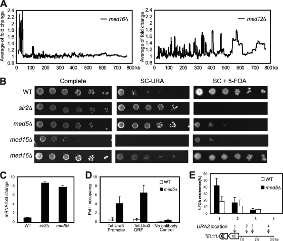
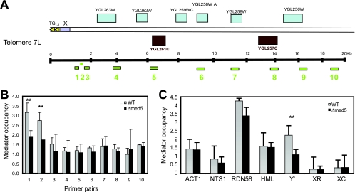
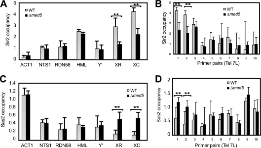
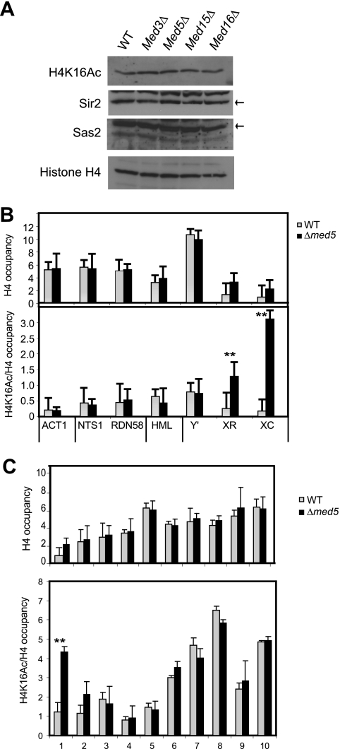
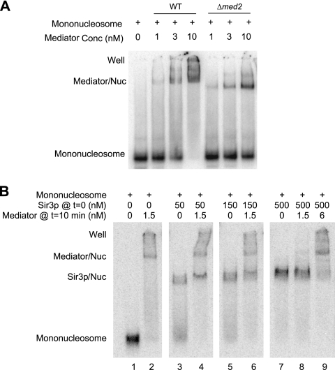
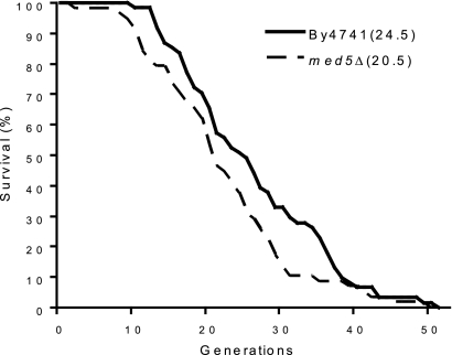
The Mediator complex is required for the regulated transcription of nearly all RNA polymerase II-dependent genes. Here we demonstrate a new role for Mediator which appears to be separate from its function as a transcriptional coactivator. Mediator associates directly with heterochromatin at telomeres and influences the exact boundary between active and inactive chromatin. Loss of the Mediator Med5 subunit or mutations in Med7 cause a depletion of the complex from regions located near subtelomeric X elements, which leads to a change in the balance between the Sir2 and Sas2 proteins. These changes in turn result in increased levels of H4K16 acetylation near telomeres and in desilencing of subtelomeric genes. Increases in H4K16 acetylation have been observed at telomeres in aging cells. In agreement with this observation, we found that the loss of MED5 leads to shortening of the Saccharomyces cerevisiae (budding yeast) replicative life span.


Figures

Fig. 1.
Fig. 1.
Deletion of Mediator components disrupts transcription silencing at telomeres. (A) Moving average analysis of gene expression changes in med16Δ and med12Δ mutant strains. The distance from a gene to the telomere was calculated based on the position of the translation start site. The window size is 150. (B) Fivefold dilutions of the indicated deletion strains were spotted on complete medium, on synthetic complete medium lacking uracil (SC−URA), and on synthetic complete medium containing 5-FOA (SC + 5-FOA). All strains (UCC3505, CGC209, CGC210, CGC211, and CGC212) contained the URA3 reporter gene inserted at tel7L. (C) Real-time PCR analysis shows fold changes of tel7L URA3 gene transcription in strains UCC3505, CGC210, and CGC209. (D) A ChIP assay using the 4H8 antibody followed by real-time PCR analysis shows Pol II occupancy at URA3 promoter and coding regions of the indicated strains. Background values were calculated using a no-antibody control. Error bars indicate standard deviations based on experiments from at least three independent cultures. (E) The X core elements of telomeres show silencing defects in med5Δ cells. The positions (1 to 4) of URA3 gene insertions near tel11L are indicated. Silencing assays were performed with wild-type and med5Δ cells bearing the URA3 gene inserted at positions 1 to 4. The extent of silencing is expressed as the fraction of cells resistant to 5-FOA (n = 4). Error bars shows standard deviations. (Schematic diagram adapted from reference with permission of the publisher.)
Fig. 2.
Fig. 2.
Mediator complex occupancy near telomeres. (A) Schematic diagram of primer pairs detecting positions at various distances (0.9 kb to 20 kb) from tel7L. (B) Mediator occupancy at tel7L. ChIP analyses were performed with material from wt (BY4741) and med5Δ mutant (CGC117) strains. Mediator occupancy was monitored using an antibody against the Med1 protein, followed by real-time PCR quantification. Error bars indicate standard deviations for at least three independent cultures. (C) Mediator occupancy at different genomic loci, including euchromatin (ACT1), ribosomal DNA (NTS1 and RDN58), a mating type locus (HML-α1), and subtelomeric regions (the X and Y′ elements of telomere 5R). X elements include X core (XC) positions and X repeat (XR) positions. The analysis was performed as described for panel B. **, P < 0.05.
Fig. 3.
Fig. 3.
Sir2 and Sas2 protein occupancy changes in med5Δ mutant cells. (A and B) Sir2 occupancy was determined by ChIP assays with wt (BY4741) and med5Δ (CGC117) mutant cells, using the primers shown in Fig. 2. (C and D) Sas2 occupancy was determined as described for panels A and B. Data shown are averages for at least three independent experiments. Error bars represent standard deviations. **, P < 0.05.
Fig. 4.
Fig. 4.
H4K16 acetylation levels change in med5Δ mutant cells. (A) Western analyses revealed that the overall levels of Sir2, Sas2, H4K16 acetylation, and H4 remained unchanged in a med5Δ strain (CGC117) compared to a wt strain (BY4741). The Sir2 and Sas2 proteins are indicated with black arrows. (B) ChIP analysis of histone H4 levels and H4K16 acetylation in med5Δ and wt cells. The genomic locations are indicated in Fig. 2. (C) ChIP analysis of histone H4 and H4K16 acetylation levels near telomere 7L. H4K16 acetylation and histone H4 occupancy were normalized to the input. H4K16 acetylation was also normalized to the histone H4 level. Error bars indicate standard deviations calculated for at least three independent cultures. **, P < 0.05.
Fig. 5.
Fig. 5.
The Mediator complex and the Sir3 protein compete for binding to mononucleosomes. Electrophoretic mobility shift assays investigated the binding of wild-type and mutant Mediator complexes to 32P-labeled mononucleosomes. Each panel shows the result of an individual experiment where all samples were run in the same gel. Mediator/Nuc, Mediator-mononucleosome cocomplex; Sir3p/Nuc, Sir3-mononucleosome cocomplex; well, material that was unable to enter the gel. (A) Wild-type and Δmed2 Mediator complexes bind to mononucleosomes with comparable affinities. Different concentrations of Mediator were incubated with an invariant concentration of mononucleosomes (0.16 nM), and the reaction mixture was loaded onto the gel. (B) Various concentrations of Sir3 (as shown in the figure) were incubated with 32P-labeled reconstituted mononucleosomes (0.16 nM) for 10 min, after which Mediator (at the concentrations specified in the figure) was added to the binding reaction mixtures, followed by incubation for another 10 min.
Fig. 6.
Fig. 6.
The med5Δ strain displays a shorter replicative life span than that of the wild-type strain (for BY4741, median replicative life span = 24.5 generations; for med5Δ strain, median replicative life span = 20.5 generations; P = 0.029). Life spans were determined by counting the total number of daughters generated by each mother cell. Experiments were performed with 64 virgin cells per plate.

References

    1. Andrau J. C., et al. 2006. Genome-wide location of the coactivator mediator: binding without activation and transient Cdk8 interaction on DNA. Mol. Cell 22: 179–192 - PubMed
    1. Askree S. H., et al. 2004. A genome-wide screen for Saccharomyces cerevisiae deletion mutants that affect telomere length. Proc. Natl. Acad. Sci. U. S. A. 101: 8658–8663 - PMC - PubMed
    1. Baidoobonso S. M., Guidi B. W., Myers L. C. 2007. Med19(Rox3) regulates intermodule interactions in the Saccharomyces cerevisiae mediator complex. J. Biol. Chem. 282: 5551–5559 - PubMed
    1. Beve J., et al. 2005. The structural and functional role of Med5 in the yeast Mediator tail module. J. Biol. Chem. 280: 41366–41372 - PubMed
    1. Bjorklund S., Gustafsson C. M. 2004. The mediator complex. Adv. Protein Chem. 67: 43–65 - PubMed

Publication types

MeSH terms

LinkOut - more resources