. 2011 Jun 9;117(23):6383-91.
doi: 10.1182/blood-2010-10-313072. Epub 2011 Apr 11.

Endothelial cell activation by antiphospholipid antibodies is modulated by Kruppel-like transcription factors

Affiliations

Endothelial cell activation by antiphospholipid antibodies is modulated by Kruppel-like transcription factors

Kristi L Allen et al. Blood. .

Abstract

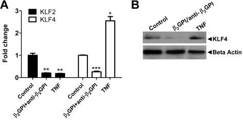
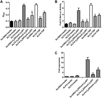
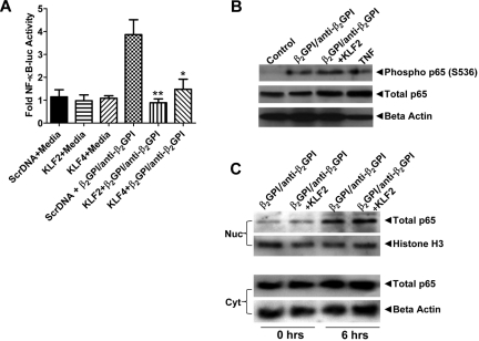
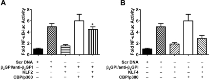
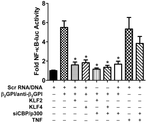
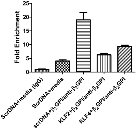
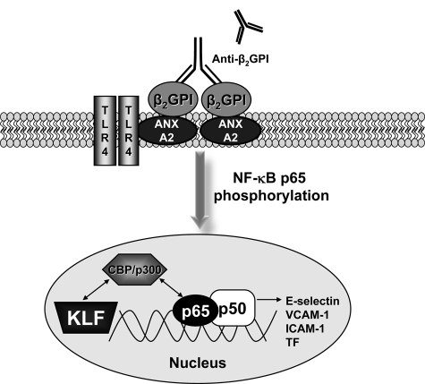
Antiphospholipid syndrome is characterized by thrombosis and/or recurrent pregnancy loss in the presence of antiphospholipid antibodies (APLAs). The majority of APLAs are directed against phospholipid-binding proteins, particularly β₂-glycoprotein I (β₂GPI). Anti-β₂GPI antibodies activate endothelial cells in a β₂GPI-dependent manner through a pathway that involves NF-κB. Krüppel-like factors (KLFs) play a critical role in regulating the endothelial response to inflammatory stimuli. We hypothesized that activation of endothelial cells by APLA/anti-β₂GPI antibodies might be associated with decreased expression of KLFs, which in turn might facilitate cellular activation mediated through NF-κB. Our experimental results confirmed this hypothesis, demonstrating markedly decreased expression of KLF2 and KLF4 after incubation of cells with APLA/anti-β₂GPI antibodies. Restoration of KLF2 or KLF4 levels inhibited NF-κB transcriptional activity and blocked APLA/anti-β₂GPI-mediated endothelial activation despite NF-κB p65 phosphorylation. Chromatin immunoprecipitation analysis demonstrated that inhibition of NF-κB transcriptional activity by KLFs reflects sequestration of the cotranscriptional activator CBP/p300, making this cofactor unavailable to NF-κB. These findings suggest that the endothelial response to APLA/anti-β₂GPI antibodies reflects competition between KLFs and NF-κB for their common cofactor, CBP/p300. Taken together, these observations are the first to implicate the KLFs as novel participants in the endothelial proinflammatory response to APLA/anti-β₂GPI antibodies.


Figures

Figure 1
Figure 1
Endothelial cell activation induced by APLA/anti-β2GPI antibodies decreases expression of KLF2 and KLF4. (A) Cells were incubated with medium alone (control), β2GPI (100nM)/anti-β2GPI antibodies (600nM), or TNF-α (10 ng/mL) for 5 hours. Total RNA was then isolated and KLF2 and KLF4 expression analyzed by quantitative real-time PCR. β2GPI/anti-β2GPI antibodies and TNF-α reduced KLF2 mRNA 5-fold compared with control (**P < .001 for both), whereas KLF4 mRNA levels were reduced 3.8-fold in the presence of β2GPI/anti-β2GPI antibodies (***P < .0001) but increased 2.6-fold by TNF-α (*P = .0012). Error bars represent the mean ± SEM of triplicate points. (B) Immunoblotting for KLF4 protein. Cells were treated as in panel A. Extracts were prepared and total protein (80 μg) separated using 7.5% SDS-PAGE, transferred to PVDF, and probed with goat anti–human KLF4 antibodies.
Figure 2
Figure 2
Restoration of KLF2 or KLF4 protects endothelial cells from APLA-mediated activation. (A) Endothelial cells were transfected with expression vectors containing scrambled DNA (Scr DNA, control), or KLF2 or KLF4 cDNA. Transfected cells were then incubated with medium alone, β2GPI (100nM) and anti-β2GPI antibodies (600nM), or TNF-α (10 ng/mL) for 5 hours, after which endothelial cell activation was measured using an E-selectin enzyme-linked immunosorbent assay. Activation was blocked in cells transfected with KLF2 or KLF4, but not scrambled DNA (**P < .01 and *P < 0.05 for KLF2 or KLF4, respectively, vs control). KLF2 and KLF 4 also blocked cellular activation in response to TNF-α. Error bars represent the mean ± SEM of quadruplicate points, and data are representative of 4 experiments. (B) Cells were treated as in panel A but cotransfected with an E-selectin-luciferase reporter and Renilla luciferase (to control for transfection efficiency). Endothelial cell activation was measured as a function of E-selectin luciferase activity normalized to Renilla. KLF2 and KLF4 inhibited E-selectin transcription compared with cells transfected with the control vector (*P < .02 for each) Error bars represent the mean ± SEM of triplicate points, and data are representative of 3 experiments. (C) Cells were transfected with KLF expression plasmids as described in panel A and incubated with medium alone (control) or β2GPI and anti–human β2GPI antibodies for 5 hours. Total RNA was then isolated, and E-selectin mRNA expression was analyzed by quantitative real-time PCR. Expression of KLF2 or KLF4 reduced the induction of E-selectin mRNA expression in response to β2GPI and anti-β2GPI antibodies by 6.7- and 2.6-fold, respectively (**P < .001 and *P < 0.01, respectively). Error bars represent the mean ± SEM of triplicate points.
Figure 3
Figure 3
Expression of KLF2 or KLF4 inhibits NF-κB transcriptional activity in APLA/anti-β2GPI–treated endothelial cells. (A) Endothelial cells were transfected with expression vectors containing scrambled DNA (Scr DNA, control), or KLF2 or KLF4 cDNA along with an NF-κB–luciferase reporter and Renilla luciferase (as a transfection efficiency control). Transfected cells were subsequently incubated with medium alone, β2GPI (100nM) and anti-β2GPI antibodies (600nM), or TNF-α (10 ng/mL) for 5 hours before measurement of luciferase activity. NF-κB transcriptional activity was determined from measured NF-κB luciferase activity normalized to Renilla. Expression of KLF2 and KLF4 inhibited NF-κB transcriptional activity in the presence of β2GPI/anti-β2GPI antibodies compared with control cells (**P < .004 and *P < 0.04, respectively). Error bars represent the mean ± SEM of triplicate points, and data are representative of 4 experiments. (B) KLF2 expression does not inhibit phosphorylation of p65 serine 536. Endothelial cells were transfected with expression vectors containing scrambled DNA (Scr DNA, control), or KLF2 cDNA and subsequently treated as in panel A. Cell extracts were prepared, and 80 μg of total protein was separated using 7.5% SDS-PAGE, transferred to PVDF, and blotted with rabbit anti–human antibodies to phospho-p65 (serine 536) and total p65. (C) KLF2 expression does not block nuclear translocation of NF-κB p65. Endothelial cells were transfected and treated as in panel B. Nuclear (Nuc) and cytoplasmic (Cyt) extracts were prepared, and 40 μg of protein from each was separated by 7.5% SDS-PAGE, transferred to PVDF, and blotted with rabbit anti–human p65. Transfection of endothelial cells with a KLF4 expression vector yielded identical results as seen with KLF2 in panels B and C.
Figure 4
Figure 4
Coexpression of CBP/p300 restores NF-κB transcriptional activity in APLA/anti-β2GPI–activated endothelial cells in the presence of KLF2 or KLF4. (A) Endothelial cells were transfected with expression vectors containing scrambled DNA (Scr DNA, control) or KLF2, and/or CBP/p300. All cells were also transfected with an NF-κB–luciferase reporter and Renilla luciferase. Cells were subsequently treated with medium alone, β2GPI (100nM)/anti-β2GPI antibodies (600nM), or TNF-α (10 ng/mL) for 5 hours before measurement of NF-κB luciferase activity, which was normalized to Renilla. KLF2 expression inhibited NF-κB transcriptional activity in the presence of β2GPI/anti-β2GPI antibodies, although inhibition was reversed by CBP/p300 (*P < .005) in the presence of CBP/p300 compared with KLF2 + β2GPI/anti-β2GPI alone. Error bars represent the mean ± SEM of triplicate points, and data are representative of 3 independent experiments. (B) Endothelial cells were transfected with expression vectors containing scrambled DNA (Scr DNA, control) or KLF4, and/or CBP/p300. All cells were also transfected with an NF-κB–luciferase reporter and Renilla luciferase. KLF4 expression inhibited NF-κB transcriptional activity in the presence of β2GPI and anti-β2GPI antibodies; inhibition was partially reversed by CBP/p300 (P = .056). Error bars represent the mean ± SEM of triplicate points, and data are representative of 3 independent experiments.
Figure 5
Figure 5
Inhibition of NF-κB activity in APLA/anti-β2GPI–treated HUVECs by KLF2 or KLF4 is in part the result of CBP/p300. Endothelial cells were transfected with scrambled DNA (Scr DNA, control), or KLF2 or KLF4, in the absence or presence of siRNA to CBP/p300. All cells were also transfected with an NF-κB–luciferase reporter and Renilla luciferase. Cells were subsequently incubated with either medium alone, β2GPI (100nM), and anti–human β2GPI antibodies (600nM), or TNF (10 ng/mL) for 5 hours, then lysed before determination of NF-κB–dependent luciferase activity. KLF2 and KLF4 expression, in the absence of siCBP/p300, inhibited NF-κB transcriptional activation in the presence of β2GPI and anti-β2GPI antibodies compared with cells transfected with the control vector (*P < .008). siCBP/p300 inhibited NF-κB transcriptional activity independently, as well as in the presence of KLF2 or KLF4 (*P < .008). However, siCBP/p300 did not significantly affect NF-κB transcriptional activity in the presence of TNF-α. Error bars represent the mean ± SEM of quadruplicate points and data are representative of 3 independent experiments.
Figure 6
Figure 6
ChIP analysis demonstrates that KLF2 and KLF4 sequester CBP/p300 and decrease CBP/p300-NF-κB complex formation and binding to NF-κB–binding sequences in the E-selectin promoter. Cells were transfected with scrambled DNA or KLF2 or KLF4 expression vectors and then incubated with medium alone or with β2GPI and anti-β2GPI antibodies for 5 hours. After formaldehyde treatment, cell lysates were immunoprecipitated with control IgG or anti-CBP/p300 antibodies, and DNA within the immunoprecipitates was isolated and amplified using primers specific for NF-κB–binding sites in the E-selectin promoter. In cells transfected with scrambled DNA, immunoprecipitation with the CBP/p300 antibody coprecipitated DNA that amplified strongly with these primers, suggesting formation of an NF-κB–CBP/p300 complex bound to NF-κB–binding sites in the E-selectin promoter. In contrast, amplification of DNA from CBP/p300 immunoprecipitates of KLF2- or KLF4-transfected cells yielded a signal only slightly increased above the baseline obtained from control cells, suggesting decreased NF-κB–CBP/p300 complex formation and decreased binding of this complex to NF-κB–binding sites in the E-selectin promoter. Control IgG did not immunoprecipitate a sequence that could be amplified. The abundance of each CBP/p300 coprecipitated NF-κB–binding sequence was calculated as fold change relative to the amount precipitated by control IgG.
Figure 7
Figure 7
Model for KLF regulation of the activity of NF-κB in the presence of APLA/anti-β2GPI antibodies. CBP/p300 regulates the activation of endothelial cells because it is an essential cofactor for NF-κB and the KLFs. A dynamic equilibrium exists between binding of CBP/p300 to the KLFs and NF-κB. The decrease in KLF levels that occur during APLA/anti-β2GPI–induced endothelial cell activation allows the preferential association of CBP/p300 with NF-κB, thus promoting the transcription of NF-κB–dependent genes, such as cell surface adhesion molecules (E-selectin) and tissue factor.

References

    1. Arnout J, Vermylen J. Current status and implications of autoimmune antiphospholipid antibodies in relation to thrombotic disease. J Thromb Haemost. 2003;1(5):931–942. - PubMed
    1. Galli M, Barbui T. Antiphospholipid antibodies and thrombosis: strength of association. Hematol J. 2003;4(3):180–186. - PubMed
    1. Rand JH. The antiphospholipid syndrome. Annu Rev Med. 2003;54:409–424. - PubMed
    1. Giannakopoulos B, Passam F, Rahgozar S, Krilis SA. Current concepts on the pathogenesis of the antiphospholipid syndrome. Blood. 2007;109(2):422–430. - PubMed
    1. de Laat B, Eckmann CM, van Schagen M, Meijer AB, Mertens K, van Mourik JA. Correlation between the potency of a beta2-glycoprotein I-dependent lupus anticoagulant and the level of resistance to activated protein C. Blood Coagul Fibrinolysis. 2008;19(8):757–764. - PubMed

Publication types

MeSH terms