. 2011 Apr 18;193(2):307-18.
doi: 10.1083/jcb.201012093. Epub 2011 Apr 11.

Defective nucleotide excision repair with normal centrosome structures and functions in the absence of all vertebrate centrins

Affiliations

Defective nucleotide excision repair with normal centrosome structures and functions in the absence of all vertebrate centrins

Tiago J Dantas et al. J Cell Biol. .

Abstract

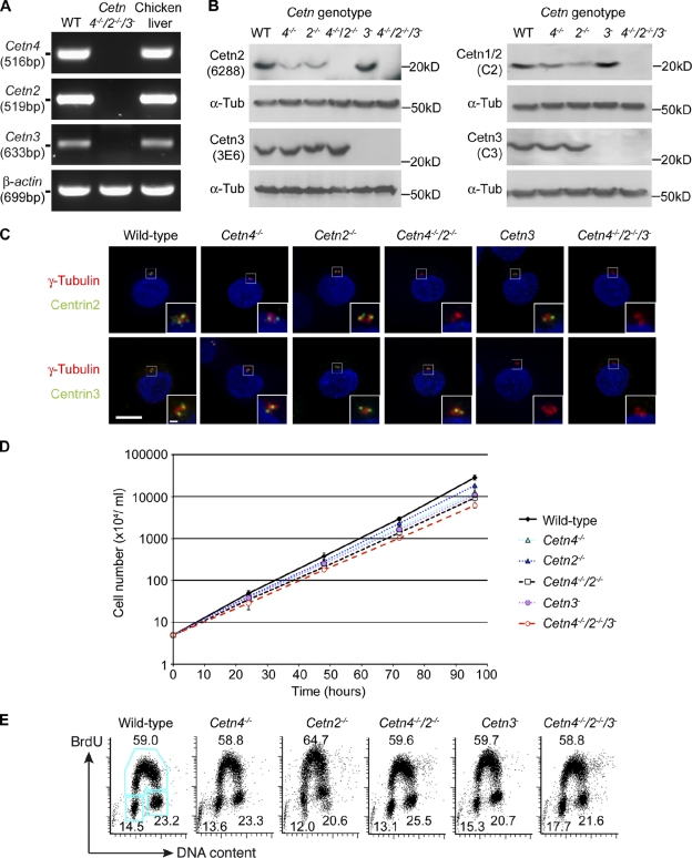
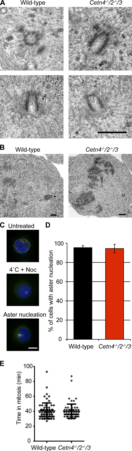
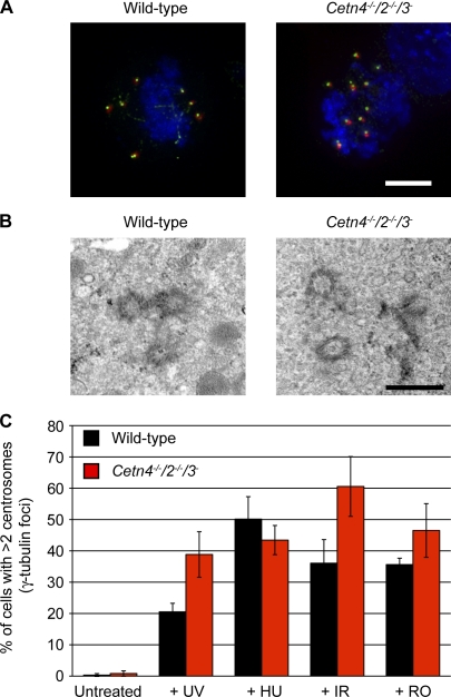
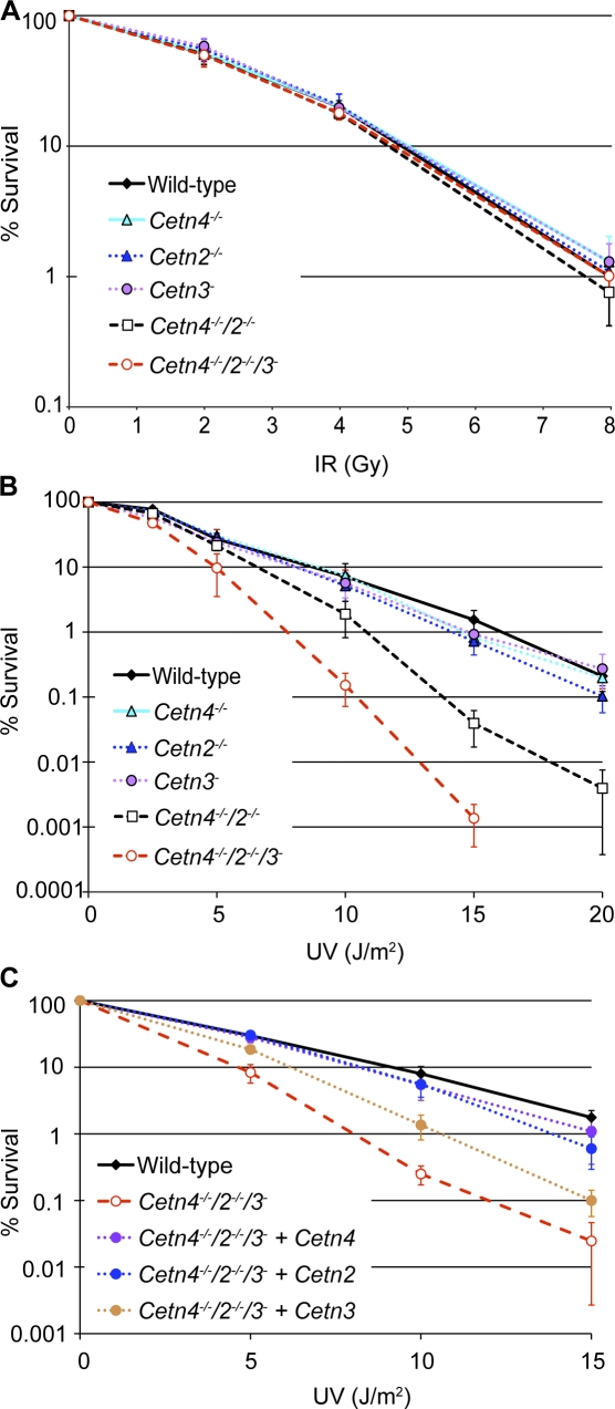
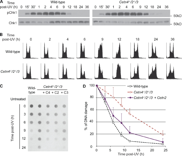
The principal microtubule-organizing center in animal cells, the centrosome, contains centrin, a small, conserved calcium-binding protein unique to eukaryotes. Several centrin isoforms exist and have been implicated in various cellular processes including nuclear export and deoxyribonucleic acid (DNA) repair. Although centrins are required for centriole/basal body duplication in lower eukaryotes, centrin functions in vertebrate centrosome duplication are less clear. To define these roles, we used gene targeting in the hyperrecombinogenic chicken DT40 cell line to delete all three centrin genes in individual clones. Unexpectedly, centrin-deficient cells underwent normal cellular division with no detectable cell cycle defects. Light and electron microscopy analyses revealed no significant difference in centrosome composition or ultrastructure. However, centrin deficiency made DT40 cells highly sensitive to ultraviolet (UV) irradiation, with Cetn3 deficiency exacerbating the sensitivity of Cetn4/Cetn2 double mutants. DNA damage checkpoints were intact, but repair of UV-induced DNA damage was delayed in centrin nulls. These data demonstrate a role for vertebrate centrin in nucleotide excision repair.


Figures

Figure 1.
Figure 1.
Centrin-deficient DT40 cells are viable and show no proliferative defect. (A) RT-PCR analysis of expression of the indicated genes in wild-type (WT) and centrin-deficient DT40 cells and in chicken liver. (B) Immunoblot of centrin in cells of the indicated genotype. Numbers refer to the centrin mutant in each lane. Antibodies used for these analyses are indicated. (C) Immunofluorescence microscopy analysis of wild-type and centrin-targeted DT40 cells stained with antibodies to the indicated centrin orthologue (green) with γ-tubulin (red) as a reference marker. Cells were counterstained with DAPI to visualize the DNA (blue) before imaging. Insets show magnifications as indicated in the main images. Bars: 5 µm; (inset) 0.5 µm. (D) Proliferation analysis of cells of the indicated genotype. Data points show mean ± SD of the results from at least three separate experiments. Doubling times were 7 h 42 min ± 28 min for wild type and 9 h 18 min ± 13 min for Cetn4/2/3-deficient cells. (E) FACS plot of cell cycle distributions in asynchronous cells of the indicated genotype. The G1 (bottom left), S (top), and G2/M (bottom right) gates are indicated in blue, and the numbers refer to the percentage of cells detected in each of the gates averaged from two separate experiments.
Figure 2.
Figure 2.
Structural integrity of centrin-deficient centrosomes. (A) Immunofluorescence microscopy analysis of wild-type and the indicated Cetn-targeted DT40 cells stained with antibodies to the indicated centrosome component (green) with γ-tubulin (red) as a reference marker. Cells were counterstained with DAPI to visualize the DNA (blue) before imaging. Bar, 5 µm. (B) Immunofluorescence micrograph of centrosomes in mitotic cells of the indicated genotype stained as in A but with α-tubulin (red) as the control. Bar, 5 µm. (C) Transmission electron micrographs of centrosomes in cells of the indicated genotype. Bars, 100 nm. (D) TEM comparison of different stages of centrosome cycle in wild-type and centrin-deficient cells. Bars, 100 nm.
Figure 3.
Figure 3.
Normal microtubule nucleation functions of centrin-deficient centrosomes. (A) Transmission electron micrographs of microtubule anchorage at the centrosomes in cells of the indicated genotype. Bar, 500 nm. (B) Transmission electron micrographs of spindle microtubule association with chromosomes in cells of the indicated genotype. Bars, 500 nm. (C) Immunofluorescence microscopy analysis of microtubule nucleation in DT40 cells before and after release from 1-h arrest in 1 µg/ml nocodazole at 4°C. Cells were stained with antibodies to α-tubulin (green) with γ-tubulin (red) as a centrosomal marker. Cells were counterstained with DAPI to visualize the DNA (blue) before imaging. Bar, 5 µm. (D) Quantitation of the percentage of cells with aster nucleation after microtubule regrowth for 1 min at 39.5°C. Histogram shows mean ± SD of three separate experiments in which at least 200 cells per experiment were counted. (E) Duration of mitosis in centrin-deficient cells. Cells of the indicated genotypes that stably expressed histone H2B-RFP were imaged by time-lapse microscopy, and the time taken from chromosome condensation to decondensation was assessed. Data show mean ± SD for the 70 individual cells analyzed as data points.
Figure 4.
Figure 4.
Efficient DNA damage–induced centrosome amplification in centrin-deficient cells. (A) Immunofluorescence microscopy analysis showing centrosome amplification in wild-type and the indicated Cetn-targeted DT40 cells stained with antibodies to glutamylated tubulin (green) and γ-tubulin (red) at 24 h after treatment with 10 Gy IR. Cells were counterstained with DAPI to visualize the DNA (blue) before imaging. Bar, 5 µm. (B) TEM comparison of amplified centrosomes in wild-type and centrin-deficient cells, seen 24 h after 10 Gy IR. Bar, 500 nm. (C) Quantitation of cells of the indicated genotype with aberrant centrosome numbers 24 h after 5-J/m2 UV-C treatment (+UV) or 10 Gy IR (+IR) and after 24-h incubation with 4 mM HU (+HU) or 6 µM RO3306 (+RO). Histogram shows mean ± SD of three separate experiments in which at least 300 cells per experiment were counted.
Figure 5.
Figure 5.
Hypersensitivity to UV irradiation of centrin-deficient cells and rescue by expression of centrins 4, 2, or 3. (A) Clonogenic survival assay of cells of the indicated genotype treated with the indicated doses of IR. Data points show mean ± SD of the surviving fractions in at least three separate experiments. (B) Clonogenic survival assay of cells of the indicated genotype treated with the indicated doses of UV-C irradiation. Data points show mean ± SD of the surviving fractions in at least three separate experiments. (C) Cetn4/2/3-deficient cells that were stably transfected with the indicated myc-centrin–expressing transgenes were analyzed by clonogenic survival assay. Survival curves show mean ± SD of the surviving fractions in at least three separate experiments.
Figure 6.
Figure 6.
Defective NER in centrin-deficient cells. (A) Immunoblot analysis of Chk1 phosphorylation in cells of the indicated genotype was performed before or at different times after 3-J/m2 UV-C treatment, using anti–phospho-Chk1 S345 with monoclonal mouse anti-Chk1 as loading control. (B) Flow cytometry analysis of the DNA content of wild-type and Cetn4/2/3-deficient cells before or at the indicated times after 3-J/m2 UV-C treatment. (C) Immunodot-blot analysis of CPDs in genomic DNA from wild type, Cetn4/2/3-deficient cells, and Cetn4/2/3-deficient cells that were stably transfected with the indicated centrin-expressing transgenes (C4, C2, and C3) before or at the indicated times after 5-J/m2 UV-C treatment. Time 0 is immediately after irradiation. (D) Quantitation of the kinetics of CPD repair in cells of the indicated genotype after 5-J/m2 UV-C treatment. Data show mean ± SD of the CPD signal normalized to time 0 signals in at least three separate experiments in which each sample was blotted in triplicate.

Similar articles

Cited by

References

    1. Acu I.D., Liu T., Suino-Powell K., Mooney S.M., D’Assoro A.B., Rowland N., Muotri A.R., Correa R.G., Niu Y., Kumar R., Salisbury J.L. 2010. Coordination of centrosome homeostasis and DNA repair is intact in MCF-7 and disrupted in MDA-MB 231 breast cancer cells. Cancer Res. 70:3320–3328 10.1158/0008-5472.CAN-09-3800 - DOI - PMC - PubMed
    1. Alieva I.B., Vorobjev I.A. 2004. Vertebrate primary cilia: a sensory part of centrosomal complex in tissue cells, but a “sleeping beauty” in cultured cells? Cell Biol. Int. 28:139–150 10.1016/j.cellbi.2003.11.013 - DOI - PubMed
    1. Arakawa H., Lodygin D., Buerstedde J.M. 2001. Mutant loxP vectors for selectable marker recycle and conditional knock-outs. BMC Biotechnol. 1:7 10.1186/1472-6750-1-7 - DOI - PMC - PubMed
    1. Araki M., Masutani C., Takemura M., Uchida A., Sugasawa K., Kondoh J., Ohkuma Y., Hanaoka F. 2001. Centrosome protein centrin 2/caltractin 1 is part of the xeroderma pigmentosum group C complex that initiates global genome nucleotide excision repair. J. Biol. Chem. 276:18665–18672 10.1074/jbc.M100855200 - DOI - PubMed
    1. Balczon R., Bao L., Zimmer W.E., Brown K., Zinkowski R.P., Brinkley B.R. 1995. Dissociation of centrosome replication events from cycles of DNA synthesis and mitotic division in hydroxyurea-arrested Chinese hamster ovary cells. J. Cell Biol. 130:105–115 10.1083/jcb.130.1.105 - DOI - PMC - PubMed

Publication types