Review
. 2011 Apr;24(2):377-410.
doi: 10.1128/CMR.00051-10.

Epidemiology and infectivity of Plasmodium falciparum and Plasmodium vivax gametocytes in relation to malaria control and elimination

Affiliations
Review

Epidemiology and infectivity of Plasmodium falciparum and Plasmodium vivax gametocytes in relation to malaria control and elimination

Teun Bousema et al. Clin Microbiol Rev. 2011 Apr.

Abstract

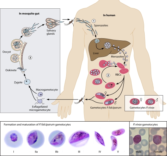
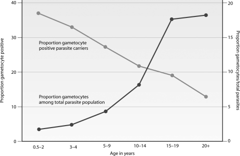
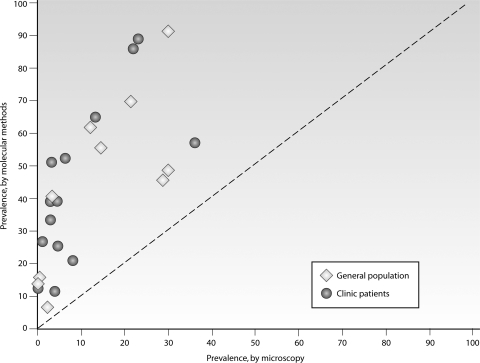
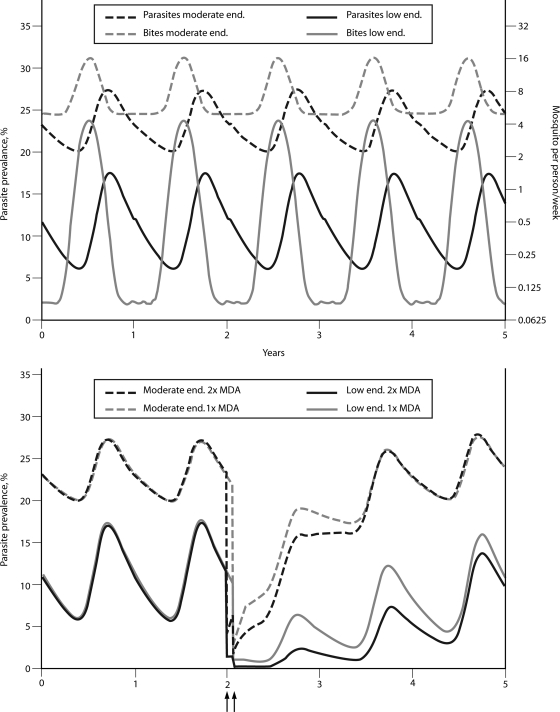
Malaria remains a major cause of morbidity and mortality in the tropics, with Plasmodium falciparum responsible for the majority of the disease burden and P. vivax being the geographically most widely distributed cause of malaria. Gametocytes are the sexual-stage parasites that infect Anopheles mosquitoes and mediate the onward transmission of the disease. Gametocytes are poorly studied despite this crucial role, but with a recent resurgence of interest in malaria elimination, the study of gametocytes is in vogue. This review highlights the current state of knowledge with regard to the development and longevity of P. falciparum and P. vivax gametocytes in the human host and the factors influencing their distribution within endemic populations. The evidence for immune responses, antimalarial drugs, and drug resistance influencing infectiousness to mosquitoes is reviewed. We discuss how the application of molecular techniques has led to the identification of submicroscopic gametocyte carriage and to a reassessment of the human infectious reservoir. These components are drawn together to show how control measures that aim to reduce malaria transmission, such as mass drug administration and a transmission-blocking vaccine, might better be deployed.


Figures

Fig. 1.
Fig. 1.
(Top) Life cycle of Plasmodium falciparum and gametocyte development. Malaria parasites enter the human bloodstream in the form of sporozoites that are injected by infected female Anopheles mosquitoes taking a blood meal. The majority of sporozoites migrate to the liver, where they invade hepatocytes and multiply. Merozoites are formed that are released into the bloodstream, where they invade red blood cells, initiating the asexual multiplication cycle. A fraction of merozoites that are released from infected red blood cells form gametocytes, the transmissible parasite form. The formation and maturation of gametocytes take place in five morphologically recognizable stages. Early-stage gametocytes are sequestered, and only mature stage V gametocytes circulate in the peripheral blood, where they can be taken up by mosquitoes. Once ingested by mosquitoes, each individual gametocyte forms 1 female macrogamete or up to 8 male microgametes. In the mosquito midgut, the fusion of gametes results in the formation of a zygote that develops into a motile ookinete that can penetrate the midgut wall to form oocysts. The oocysts enlarge over time and burst to release sporozoites that migrate to the mosquito salivary gland, rendering the mosquito infectious to human beings. (Bottom) The five developmental stages of P. falciparum gametocytes and mature P. vivax gametocytes. (The P. falciparum gametocyte photographs are reprinted from reference with permission; the P. vivax gametocyte photographs are courtesy of Debbie Nolder, Malaria Reference Laboratory, London School of Hygiene and Tropical Medicine, United Kingdom, reproduced with permission.)
Fig. 2.
Fig. 2.
Prevalence of gametocytes in different transmission settings and age groups. Gametocytes were detected by microscopy (thick blood films; screening of 100 fields at a magnification of ×1,000, using oil immersion) in cross-sectional surveys in areas of high and seasonal transmission intensity in Burkina Faso (336) and the Gambia (121), high perennial transmission in Tanzania (121), and low seasonal transmission intensity in Tanzania (413). Gametocytes are detected in a larger proportion of the population in settings of high endemicity, where gametocyte prevalence depends on age; in settings of low endemicity, gametocyte carriage is rare and more evenly distributed across age groups.
Fig. 3.
Fig. 3.
Age-dependent carriage of gametocytes in an area of high and seasonal malaria transmission in Burkina Faso. Gametocytes were detected by microscopy (thick blood films; screening of 100 fields at a magnification of ×1,000, using oil immersion) in cross-sectional surveys. The proportion of infections with concurrent gametocytes is highest in children and decreases with age. Among infections with gametocytes, the density of gametocytes relative to the total parasite density increases with age (334).
Fig. 4.
Fig. 4.
Gametocyte carriage by microscopy and molecular detection tools. Gametocytes were detected by microscopy, typically screening 100 microscopic fields, and by pfs25- or pfg377-specific RT-PCR, LAMP, or QT-NASBA. Samples were derived from the general population in Burkina Faso, Tanzania, the Gambia, and Thailand (open diamonds) (70, 263, 319, 335, 337, 340, 413) and from people attending clinics in Kenya, Tanzania, Sudan, and Vietnam (closed circles) (1, 55, 140, 256, 279, 314, 412), mostly children participating in clinical trials. (Reproduced from reference 334 with permission.)
Fig. 5.
Fig. 5.
Gametocyte densities and mosquito infection rates. Data from two transmission studies of asymptomatic children in Burkina Faso (335) and symptomatic children 2 weeks after antimalarial treatment in Kenya (55, 402) were combined. Both studies determined gametocyte carriage by Pfs25-specific QT-NASBA and offered venous blood to mosquitoes in a membrane feeding assay (see “Evidence for Naturally Acquired Transmission-Blocking Activity”) to determine the proportion of infected mosquitoes. Circles indicate aggregated data, grouped in 10 categories of similar gametocyte densities.
Fig. 6.
Fig. 6.
Gametocytes as an early indicator of parasite resistance. The data presented are from a study in the Gambia where children were treated with chloroquine (184). The figure presents gametocyte prevalence by microscopy (thick blood films; screening of 100 high-power fields at a magnification of ×1,000, using oil immersion) on day 7 after treatment (left y axis; error bars indicate the upper limit of the 95% confidence interval), median gametocyte density by microscopy in gametocyte carriers on day 7 after treatment (right y axis; error bars indicate the upper limit of the interquartile range), and the mean proportion of infected mosquitoes determined by membrane feeding assays (right y axis; error bars indicate the upper limit of the 95% confidence interval). Wild type, no mutations detected at enrollment in two genes related to chloroquine resistance, P. falciparum multidrug resistance gene 1 (Pfmdr1 86Y) and P. falciparum chloroquine resistance transporter (Pfcrt 76T); single mutant, mutation detected in either Pfmdr1 or Pfcrt; double mutant, mutation detected in both Pfmdr1 and Pfcrt.
Fig. 7.
Fig. 7.
Three different experimental setups for assessing malaria transmission and TRA. In direct skin feeding assays, reared (malaria-free) mosquitoes are allowed to feed directly on blood through the skin of a naturally infected individual. Oocysts can be detected on the mosquito midgut after dissection of the mosquito. This is mostly done 7 to 9 days after feeding. The presence of oocysts confirms infection; TRA cannot be determined by direct skin feeding. In the direct membrane feeding assay (DMFA), a venous blood sample is taken from a naturally infected individual. This blood sample forms the source of gametocytes and test plasma. Samples are centrifuged, and the red blood cells that contain gametocytes are subsequently mixed with autologous plasma or malaria-naïve control serum. This mixture is kept at 37°C and offered to reared mosquitoes through a membrane, commonly Parafilm. Seven days after feeding, mosquitoes are dissected. The prevalence or density of infection in mosquitoes can be compared in pairwise comparisons between the batch of mosquitoes that fed on gametocytes in combination with control serum and the batch of mosquitoes that fed on gametocytes with autologous plasma. In the standard membrane feeding assay (SMFA), cultured gametocytes are offered to mosquitoes in the presence of test or control sera. The prevalence or density of infection in batches of mosquitoes feeding on gametocytes with control serum is compared to that in batches with test serum. In the SMFA, a large number of test sera can be tested simultaneously.
Fig. 8.
Fig. 8.
Predicted impacts of mass drug administration in two settings of malaria endemicity, using two different transmission scenarios. (Top) Situation of seasonal transmission in an area of moderate endemicity and an area of low endemicity. In the area of moderate endemicity, parasite prevalence is ∼20% in the general population in the dry season, when people are exposed to ∼5 mosquito bites/person/week; in the wet season, parasite prevalence increases to ∼28% and mosquito exposure to 16 bites/person/week. In the setting of low endemicity, parasite prevalence is ∼5% in the dry season, when people are exposed to an average of one mosquito bite/2 months; in the wet season, parasite prevalence increases to ∼18% and mosquito exposure to 4 bites/person/week. (Bottom) Simulated impact of one or two rounds of MDA toward the end of the dry season. The timing of the MDA is indicated with an arrow; in the scenario where MDA is repeated, the gap between the two rounds of MDA is 1 month, and the correlation between coverage in the first and second rounds of MDA is 0.5. The drug used for MDA has the characteristics of artemether-lumefantrine, with an efficacy against asexual parasites of 95% and a duration of (submicroscopic) gametocyte carriage of 13 days after initiation of treatment. The coverage with the intervention is 95%.

References

    1. Abdel-Wahab A., et al. 2002. Dynamics of gametocytes among Plasmodium falciparum clones in natural infections in an area of highly seasonal transmission. J. Infect. Dis. 185:1838–1842 - PubMed
    1. Abu-Raddad L. J., Patnaik P., Kublin J. G. 2006. Dual infection with HIV and malaria fuels the spread of both diseases in sub-Saharan Africa. Science 314:1603–1606 - PubMed
    1. Adjuik M., et al. 2004. Artesunate combinations for treatment of malaria: meta-analysis. Lancet 363:9–17 - PubMed
    1. Akim N. I., et al. 2000. Dynamics of P. falciparum gametocytemia in symptomatic patients in an area of intense perennial transmission in Tanzania. Am. J. Trop. Med. Hyg. 63:199–203 - PubMed
    1. Alano P. 2007. Plasmodium falciparum gametocytes: still many secrets of a hidden life. Mol. Microbiol. 66:291–302 - PubMed

MeSH terms

LinkOut - more resources