Review
. 2011 Apr;24(2):411-45.
doi: 10.1128/CMR.00056-10.

Mucormycosis caused by unusual mucormycetes, non-Rhizopus, -Mucor, and -Lichtheimia species

Affiliations
Review

Mucormycosis caused by unusual mucormycetes, non-Rhizopus, -Mucor, and -Lichtheimia species

Marisa Z R Gomes et al. Clin Microbiol Rev. 2011 Apr.

Abstract

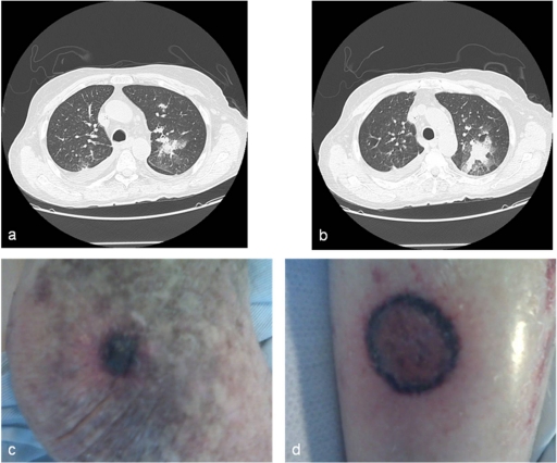
Rhizopus, Mucor, and Lichtheimia (formerly Absidia) species are the most common members of the order Mucorales that cause mucormycosis, accounting for 70 to 80% of all cases. In contrast, Cunninghamella, Apophysomyces, Saksenaea, Rhizomucor, Cokeromyces, Actinomucor, and Syncephalastrum species individually are responsible for fewer than 1 to 5% of reported cases of mucormycosis. In this review, we provide an overview of the epidemiology, clinical manifestations, diagnosis of, treatment of, and prognosis for unusual Mucormycetes infections (non-Rhizopus, -Mucor, and -Lichtheimia species). The infections caused by these less frequent members of the order Mucorales frequently differ in their epidemiology, geographic distribution, and disease manifestations. Cunninghamella bertholletiae and Rhizomucor pusillus affect primarily immunocompromised hosts, mostly resulting from spore inhalation, causing pulmonary and disseminated infections with high mortality rates. R. pusillus infections are nosocomial or health care related in a large proportion of cases. While Apophysomyces elegans and Saksenaea vasiformis are occasionally responsible for infections in immunocompromised individuals, most cases are encountered in immunocompetent individuals as a result of trauma, leading to soft tissue infections with relatively low mortality rates. Increased knowledge of the epidemiology and clinical presentations of these unusual Mucormycetes infections may improve early diagnosis and treatment.


Figures

Fig. 1.
Fig. 1.
Taxonomic scheme of human-pathogenic Mucormycetes of the order Mucorales.
Fig. 2.
Fig. 2.
Disseminated C. bertholletiae infection. The images are contrast-enhanced CT images of the chest revealing progression of pulmonary lesions in the left side (a and b) and pictures of necrotic skin lesions in the left elbow (c) and right leg (d) of a 76-year-old man with acute lymphoblastic leukemia (unpublished case).
Fig. 3.
Fig. 3.
Unusual Mucormycetes. (a2, b2, c2, d2, and e to g) Lactophenol cotton blue mount preparations. (a1, b1, c1, and d1) Potato dextrose agar (PDA) medium plates. (a1) C. bertholletiae colony surface on a PDA medium plate. (a2) C. bertholletiae sporangiophores in terminal swellings called vesicles, with sporangioles (short arrow). (b1) Colony surface of R. pusillus on a PDA medium plate incubated at 30°C for 96 h. (b2) R. pusillus sporangiophores with globose sporangia. (c1) S. vasiformis colony surface on a PDA medium plate incubated at 30°C (48 h). (c2) S. vasiformis sporangiophore arising from a “foot cell”-like hyphal element (long arrow), flask-shaped sporangium, and liberated sporangiospores (short arrow). (d1) Actinomucor elegans colony surface on a PDA medium plate incubated at 30°C (96 h). (d2) Actinomucor elegans branched sporangiophores, sporangium (long arrow), columella (block arrow), and various sporangiospores (short arrow). (e) Unbranched Apophysomyces elegans sporangiophore (long arrow) with a pyriform sporangium (short arrow). (f) S. racemosum sporangiophores with merosporangia (short arrow). (g) C. recurvatus sporangiolating vesicle (short arrow) and zygospores (long arrow). Bars, 20 μm. (All photomicrographs except panel a1 courtesy of Deanna A. Sutton, reproduced with permission.)
Fig. 4.
Fig. 4.
Geographic distribution of reported cases of unusual mucormycosis. (Modified from a map that is freely available from psdGraphics.)
Fig. 5.
Fig. 5.
Rhino-orbital infection caused by Syncephalastrum racemosum in a 64-year-old woman with relapsed acute lymphoblastic leukemia after allogeneic hematopoietic stem cell transplantation (unpublished case). (a and b) Nasal endoscopy showing hemorrhagic and necrotic lesions with white cottony growth. (c) Contrast-enhanced CT scan demonstrating ethmoid sinusitis. (d) Lactophenol cotton blue mount preparation showing cylindrical or finger-like projections (arrow) surrounding a swollen vesicle typical of merosporangia of S. racemosum. (Courtesy of George M. Viola, reproduced with permission.)
Fig. 6.
Fig. 6.
Reported cases of unusual mucormycosis, according to Medline (searched on 23 December 2010).

Similar articles

Cited by

References

    1. Aarset H., Aasarod K., Bergan U., Angelsen A. 2001. Acute renal infarction in a woman with slight asthma. Nephrol. Dial. Transplant. 16:1711–1712 - PubMed
    1. Abdel-Hafez S. I., Moubasher A. H., Shoreit A. A., Ismail M. A. 1990. Fungal flora associated with combine harvester wheat and sorghum dusts from Egypt. J. Basic Microbiol. 30:467–479 - PubMed
    1. Abuali M. M., et al. 2009. Rhizomucor variabilis var. regularior and Hormographiella aspergillata infections in a leukemic bone marrow transplant recipient with refractory neutropenia. J. Clin. Microbiol. 47:4176–4179 - PMC - PubMed
    1. Ajello L., Dean D. F., Irwin R. S. 1976. The zygomycete Saksenaea vasiformis as a pathogen of humans with a critical review of the etiology of zygomycosis. Mycologia 68:52–62 - PubMed
    1. Al-Ajam M. R., Bizri A. R., Mokhbat J., Weedon J., Lutwick L. 2006. Mucormycosis in the Eastern Mediterranean: a seasonal disease. Epidemiol. Infect. 134:341–346 - PMC - PubMed

Substances