Review
. 2011 Aug 1;3(8):a004713.
doi: 10.1101/cshperspect.a004713.

Synthesis and biosynthetic trafficking of membrane lipids

Affiliations
Review

Synthesis and biosynthetic trafficking of membrane lipids

Tomas Blom et al. Cold Spring Harb Perspect Biol. .

Abstract

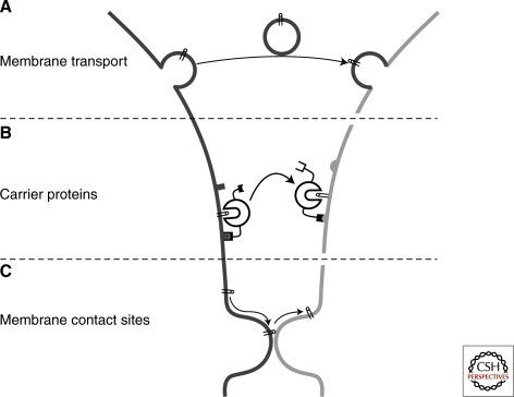
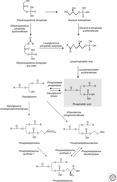
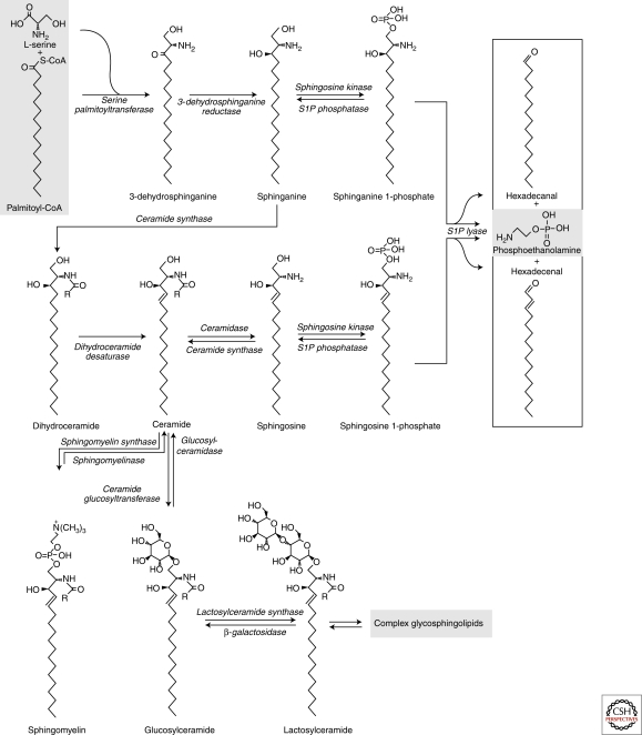
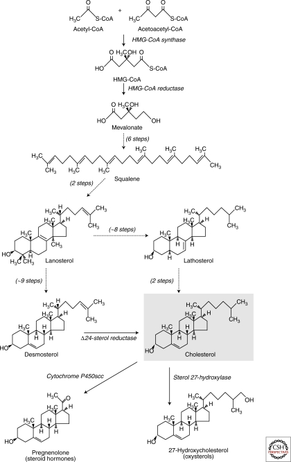
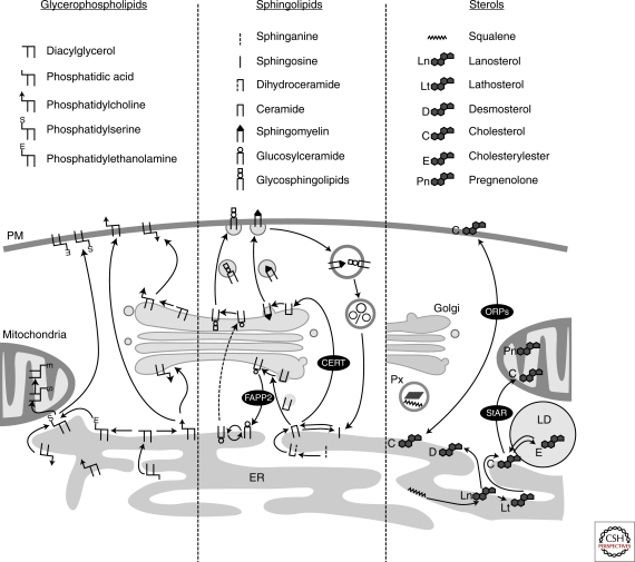
Eukaryotic cells can synthesize thousands of different lipid molecules that are incorporated into their membranes. This involves the activity of hundreds of enzymes with the task of creating lipid diversity. In addition, there are several, typically redundant, mechanisms to transport lipids from their site of synthesis to other cellular membranes. Biosynthetic lipid transport helps to ensure that each cellular compartment will have its characteristic lipid composition that supports the functions of the associated proteins. In this article, we provide an overview of the biosynthesis of the major lipid constituents of cell membranes, that is, glycerophospholipids, sphingolipids, and sterols, and discuss the mechanisms by which these newly synthesized lipids are delivered to their target membranes.


Figures

Figure 1.
Figure 1.
Mechanisms of intermembrane lipid transport. (A) Membrane transport moves lipids together with proteins in vesicular and tubular carriers that bud off from a donor membrane, and are transported along cytoskeletal tracks to the acceptor membrane, in which they fuse to deliver their cargo. (B) Cytosolic carrier proteins transfer lipids in hydrophobic pockets that show selectivity toward one or a few lipid types. Carrier proteins often contain domains that bind to the donor and/or acceptor membranes. (C) Lipid exchange may also occur between membranes that are in very close proximity. Transfer via such membrane contact sites may be facilitated by carrier proteins (combination of models B and C).
Figure 2.
Figure 2.
An overview of the biosynthetic pathways of major glycerophospholipids. De novo synthesis of the glycerophospholipids begins in the ER by a series of reduction and acylation reactions leading to the formation of phosphatidic acid. Dephosphorylation of phosphatidic acid yields diacylglycerol, which can be turned into phosphatidylcholine or phosphatidylethanolamine by the addition of a phosphocholine or a phosphoethanolamine head group, respectively. Phosphatidylserine is formed by exchanging the head group from either phosphatidylcholine or phosphatidylethanolamine for serine. Mitochondrial phosphatidylethanolamine is synthesized on location by decarboxylation of phosphatidylserine.
Figure 3.
Figure 3.
Overview of the biosynthetic pathways of sphingolipids. The sphingoid backbone is formed by the condensation of serine and palmitoyl-CoA. Three further synthetic steps are needed to produce ceramide, which the first compound with a bona fide sphingosine backbone. After its synthesis in the ER, ceramide can be metabolized into sphingomyelin or glycosphingolipids in the lumenal leaflet of the Golgi. The postceramide metabolic steps are reversible and ceramide can also be formed by the sequential degradation of more complex sphingolipids. Deacylation of ceramide yields sphingosine that can be phosphorylated to sphingosine-1-phosphate. The irreversible degradation of the sphingoid backbone is catalyzed by a lyase that acts on either sphingosine-1-phosphate or sphinganine-1-phosphate.
Figure 4.
Figure 4.
Overview of the biosynthetic pathways of sterols. The four-ring sterol backbone derives from reductive polymerizations of acetate to generate squalene, which is cyclized to form lanosterol, the first sterol in the pathway. The rate-limiting enzyme of cholesterol biosynthesis is HMG-CoA reductase. The postlanosterol pathway involves roughly 20 steps, with some of the enzymes capable of acting on multiple substrates. If the carbon-24 double bond is reduced early on, the pathway procedes via lathosterol (and 7-dehydrocholesterol, not shown) to cholesterol, whereas reduction of carbon-24 only in the last step results in the generation of desmosterol as the penultimate cholesterol cholesterol precursor. Cholesterol serves as a precursor for other bioactive sterols, such as steroid hormones and oxysterols.
Figure 5.
Figure 5.
Biosynthetic trafficking of major membrane lipids. (Left) Glycerophospholipids are synthesized in the ER, with phosphatidylserine synthases enriched in mitochondria associated membrane fractions. Glycerophospholipids are transported from the ER both along exocytic membrane transport and by nonvesicular, yet poorly characterized mechanisms. (Middle) De novo sphingolipid synthesis is initiated in the ER. The ER to Golgi transport of ceramide for the assembly of more complex sphingolipids is mediated by CERT, and to a lesser extent by membrane transport. The post-Golgi transport of complex sphingolipids is dependent on membrane transport. Sphingosine that stems from lysosomally degraded complex sphingolipids can be acylated to form ceramide in the ER and recycled to sphingomyelin and glycosphingolipids. (Right) Cholesterol biosynthetic enzymes reside in the ER, with some presqualene enzymes also localized in peroxisomes (Px). Sterols are transported to the plasma membrane (PM) largely via Golgi bypass route(s) and the ORP proteins play a role in this process, as well as in the reverse transport from the plasma membrane to the ER. Sterols imported into mitochondria by StAR can be used for steroid hormone synthesis. Excess cholesterol can be esterified in the ER by acyl-CoA cholesterol acyltransferase and stored in lipid droplets (LD). Arrows indicate the direction of lipid transport. Please note that arrows do not necessarily reflect the transport distance as membranes move constantly and lipid transfer may be facilitated by close apposition of membranes. Carrier proteins are indicated by black circles.

References

    1. Achleitner G, Gaigg B, Krasser A, Kainersdorfer E, Kohlwein SD, Perktold A, Zellnig G, Daum G 1999. Association between the endoplasmic reticulum and mitochondria of yeast facilitates interorganelle transport of phospholipids through membrane contact. Eur J Biochem 264: 545–553 - PubMed
    1. Achleitner G, Zweytick D, Trotter PJ, Voelker DR, Daum G 1995. Synthesis and intracellular transport of aminoglycerophospholipids in permeabilized cells of the yeast, Saccharomyces cerevisiae. J Biol Chem 270: 29836–29842 - PubMed
    1. Arakane F, Sugawara T, Nishino H, Liu Z, Holt JA, Pain D, Stocco DM, Miller WL, Strauss JF III 1996. Steroidogenic acute regulatory protein (StAR) retains activity in the absence of its mitochondrial import sequence: Implications for the mechanism of StAR action. Proc Natl Acad Sci 93: 13731–13736 - PMC - PubMed
    1. Baumann NA, Sullivan DP, Ohvo-Rekila H, Simonot C, Potteka A, Klaassen Z, Beh CT, Menon AK 2005. Transport of newly synthesized sterol to the sterol-enriched plasma membrane occurs via nonvesicular equilibration. Biochemistry 44: 5816–5826 - PubMed
    1. Ben-David O, Futerman AH 2010. The role of the ceramide acyl chain length in neurodegeneration: Involvement of ceramide synthases. Neuromolecular Med 12: 341–350 - PubMed

Publication types

Substances

LinkOut - more resources