. 2011 Apr 26;108(17):7058-63.
doi: 10.1073/pnas.1007293108. Epub 2011 Apr 11.

Follistatin-like 1 (Fstl1) is a bone morphogenetic protein (BMP) 4 signaling antagonist in controlling mouse lung development

Affiliations

Follistatin-like 1 (Fstl1) is a bone morphogenetic protein (BMP) 4 signaling antagonist in controlling mouse lung development

Yan Geng et al. Proc Natl Acad Sci U S A. .

Abstract

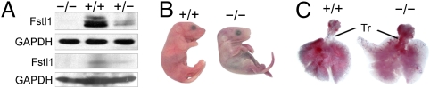
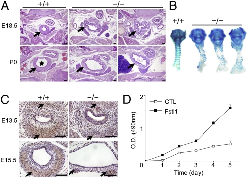
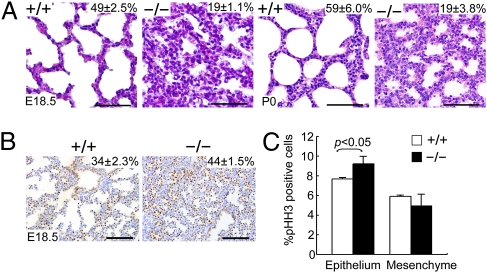
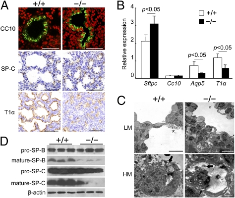
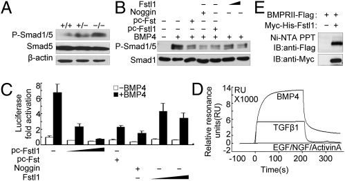
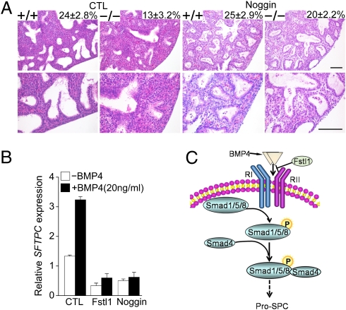
Lung morphogenesis is a well orchestrated, tightly regulated process through several molecular pathways, including TGF-β/bone morphogenetic protein (BMP) signaling. Alteration of these signaling pathways leads to lung malformation. We investigated the role of Follistatin-like 1 (Fstl1), a secreted follistatin-module-containing glycoprotein, in lung development. Deletion of Fstl1 in mice led to postnatal lethality as a result of respiratory failure. Analysis of the mutant phenotype showed that Fstl1 is essential for tracheal cartilage formation and alveolar maturation. Deletion of the Fstl1 gene resulted in malformed tracheal rings manifested as discontinued rings and reduced ring number. Fstl1-deficient mice displayed septal hypercellularity and end-expiratory atelectasis, which were associated with impaired differentiation of distal alveolar epithelial cells and insufficient production of mature surfactant proteins. Mechanistically, Fstl1 interacted directly with BMP4, negatively regulated BMP4/Smad1/5/8 signaling, and inhibited BMP4-induced surfactant gene expression. Reducing BMP signaling activity by Noggin rescued pulmonary atelectasis of Fstl1-deficient mice. Therefore, we provide in vivo and in vitro evidence to demonstrate that Fstl1 modulates lung development and alveolar maturation, in part, through BMP4 signaling.


Conflict of interest statement

The authors declare no conflict of interest.

Figures

Fig. 1.
Fig. 1.
Generation of Fstl1−/− mice. (A) Western blot analysis of Fstl1 proteins from E15.5 embryos (Upper) or E18.5 lung tissues (Lower). (B) Examination of neonates after birth revealed that Fstl1−/− neonates were cyanotic. (C) Autopsy observation showed that WT lungs were expanded by inhalation of air, which can be seen as air bubbles in the distal regions, but Fstl1−/− lungs were collapsed and did not show evidence of air in the distal airways (tr, trachea).
Fig. 2.
Fig. 2.
Tracheal malformation in Fstl1−/− mice. (A) Transverse sections of the middle portion of trachea from a WT and two homozygous mice before (E18.5) and after breath (P0) showed an increase in the lumen diameter (asterisk), dispersion and discontinuity of cartilage ring (arrow), and disorganization of the epithelial layer in the mutants. (Scale bars, 100 μm.) (B) Alcian blue staining revealed impaired banding pattern of tracheal C-ring cartilage in all mutant skeletal preparations (ventral views). (C). Sections stained with an antibody against type II collagen (arrows). (Scale bars, 100 μm.) (D). Effects of stable overexpression of Fstl1 on cell proliferation of ATDC5 cells with MTT assays.
Fig. 3.
Fig. 3.
Abnormal lung morphogenesis and lung epithelial cell hyperplasia in Fstl1−/− mice. (A) Histological analysis of embryos (E18.5) and newborn pups (P0) revealed normal inflated alveoli with thinner septa in WT, but thickened hypercellular septa with reduced airspaces in Fstl1−/−. (Scale bars, 50 μm.) (B) Distribution of TTF1 by IHC. (Scale bars, 100 μm.) The percentage of the total examined sac area of the lung section or the percentage of the TTF1-positive cells in the total cells of the lung are shown in the top right corner of each diagram (P < 0.05). (C) Quantification of cell proliferation by p-HH3 immunostaining in E15.5 WT and Fstl1−/− lungs. The graph represents the mean of four independent experiments showing the comparison of the percentages of p-HH3–positive cells in the epithelium and mesenchyme between WT and Fstl1−/− lungs.
Fig. 4.
Fig. 4.
Impaired alveolar epithelial cell differentiation/maturation in Fstl1−/− lungs. (A) Expression of differentiation marker genes for lung epithelial cells in E18.5 embryos. (Scale bars, 50 μm.) (B) The relative expression levels of differentiation marker genes in E18.5 Fstl1−/− lungs as determined by qRT-PCR. Data represent the mean ± SEM in triplicates. (C) Transmission EM of the lung septa of E18.5 embryos shown at low magnification (LM) and high magnification (HM). Squamous AEC-I cell in WT was closely opposite to densely stained capillary endothelial cell creating thin blood–air barrier (Upper Left). Cuboidal AEC-II cells in WT lungs contained many lamellar bodies (arrows) and apical microvilli (Lower Left). Surfactants (asterisk) were visible in the saccular spaces (Upper Left). The blood–air barrier was significantly thicker with increased numbers of undifferentiated cuboidal epithelial cells in Fstl1−/− (Upper Right). These cells were immature with dispersed cytoplasmic glycogen and some small lamellar bodies (arrows; Lower Right). (Scale bars: Upper, 10 μm; Lower, 2 μm.) (D) Western blotting of pro–SP-C and pro–SP-B, mature SP-C, and mature SP-B proteins in extracts of whole lungs taken from WT and Fstl1−/− embryos at E18.5.
Fig. 5.
Fig. 5.
Fstl1 modulated BMP4/Smad1/5/8 signaling via binding to BMP4. (A) Phosphorylated Smad1/5 in lung tissues from WT and Fstl1−/− embryos at E18.5. (B) Fstl1 inhibited BMP4-induced phosphorylation of Smad1/5 in Hep3B cells. Recombinant Fstl1 protein (25, 100 ng/mL) or transient overexpression of Fstl1 (pc-Fstl1, 1 μg) inhibited Smad1/5 phosphorylation induced by BMP4 (20 ng/mL). Noggin (25 ng/mL) and pc-Follistatin (Fst) (1 μg) were used as positive controls because they are known BMP antagonists. After 1 h of BMP4 treatment, cells were harvested for immunoblotting. (C) Fstl1 inhibited BMP4-induced expression of the reporter BRE-luciferase activities in Hep3B cells. The construct of BRE-luciferase reporter (0.3 μg) was cotransfected with pc-Fstl1 (0.1 μg, 0.2 μg) or treated with recombinant Fstl1 protein (25 ng/mL or 100 ng/mL). After 16 h of BMP4 treatment, cells were harvested for luciferase assay. Pc-Fst (0.1 μg) or Noggin (25 ng/mL) were used as control. The data represent the mean ± SEM of three independent experiments after normalized to Renilla activity. (D) Sensorgrams of SPR analyses show the binding of Fstl1 to BMP4 or to TGF-β1, not to activin A; EGF and NGF were used as negative controls. (E) Fstl1 binds to type II receptor of BMP4 using a pull-down assay. The Ni-Fstl1-BMPRII protein complex was immunoblotted with anti-Flag antibody to confirm the presence of BMPRII (Upper). Each blot was further developed with anti-Myc antibody to confirm the presence of Fstl1 (Lower).
Fig. 6.
Fig. 6.
Reducing BMP4 signaling activity rescued atelectasis phenotype of Fstl1−/− embryonic lung explants. (A) Noggin increased saccular dilation of Fstl1−/− fetal lung explants. E15.5 lung explants from WT and Fstl1−/− littermates were cultured for 48 to 54 h in absence (CTL) or presence of Noggin (500 ng/mL). H&E staining of explant sections shown at low (Upper) and high (Lower) magnification. Saccular dilation was shown in Noggin treated-Fstl1−/− explants. The percentage of the total examined sac area of the lung section is shown in the top right corner of each diagram (Upper). (Scale bar, 100 μm.) (B) Fstl1 (100 ng/mL) inhibited BMP4-increased SFTPC expression in A549 cells. The data represent the mean ± SEM of three independent experiments. (C) Summary of the role of Fstl1 in embryonic lung development. Fstl1 modulates BMP4-induced Smad-1/5/8 activity and inhibits pro–SP-C expression.

References

    1. Morrisey EE, Hogan BL. Preparing for the first breath: genetic and cellular mechanisms in lung development. Dev Cell. 2010;18:8–23. - PMC - PubMed
    1. Maeda Y, Davé V, Whitsett JA. Transcriptional control of lung morphogenesis. Physiol Rev. 2007;87:219–244. - PubMed
    1. Rossant J, Tam PPL. Mouse Development: Patterning, Morphogenesis, and Organogenesis. Amsterdam: Elsevier; 2002.
    1. Que J, Choi M, Ziel JW, Klingensmith J, Hogan BL. Morphogenesis of the trachea and esophagus: Current players and new roles for noggin and Bmps. Differentiation. 2006;74:422–437. - PubMed
    1. Hogan BL, Yingling JM. Epithelial/mesenchymal interactions and branching morphogenesis of the lung. Curr Opin Genet Dev. 1998;8:481–486. - PubMed

Publication types

MeSH terms

LinkOut - more resources