. 2011 Apr 26;108(17):7028-33.
doi: 10.1073/pnas.1009188108. Epub 2011 Apr 11.

Cargo binding activates myosin VIIA motor function in cells

Affiliations

Cargo binding activates myosin VIIA motor function in cells

Tsuyoshi Sakai et al. Proc Natl Acad Sci U S A. .

Abstract

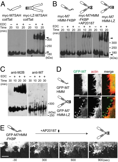
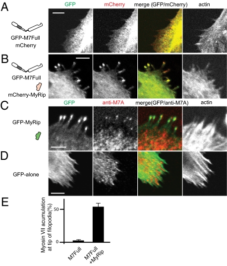
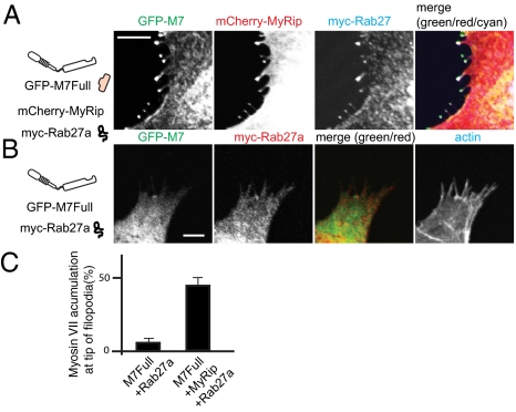
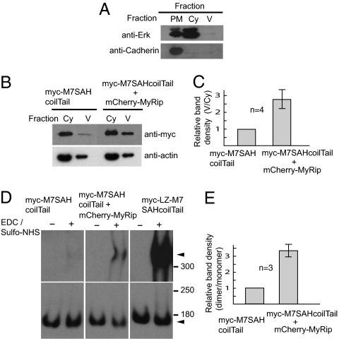
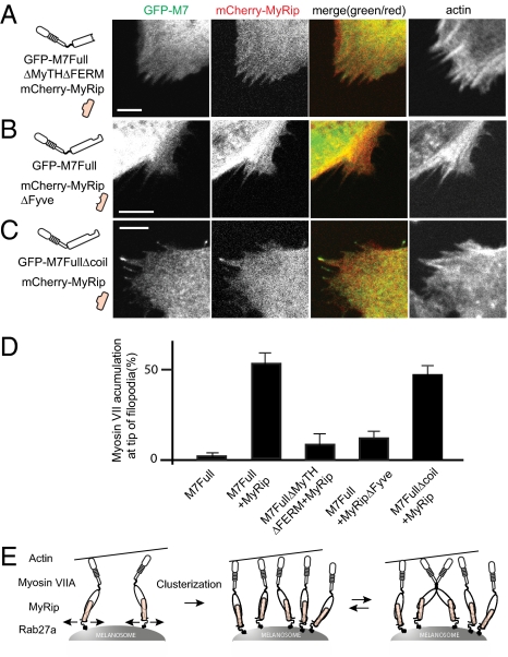
Myosin VIIA, thought to be involved in human auditory function, is a gene responsible for human Usher syndrome type 1B, which causes hearing and visual loss. Recent studies have suggested that it can move processively if it forms a dimer. Nevertheless, it exists as a monomer in vitro, unlike the well-known two-headed processive myosin Va. Here we studied the molecular mechanism, which is currently unknown, of activating myosin VIIA as a cargo-transporting motor. Human myosin VIIA was present throughout cytosol, but it moved to the tip of filopodia upon the formation of dimer induced by dimer-inducing reagent. The forced dimer of myosin VIIA translocated its cargo molecule, MyRip, to the tip of filopodia, whereas myosin VIIA without the forced dimer-forming module does not translocate to the filopodial tips. These results suggest that dimer formation of myosin VIIA is important for its cargo-transporting activity. On the other hand, myosin VIIA without the forced dimerization module became translocated to the filopodial tips in the presence of cargo complex, i.e., MyRip/Rab27a, and transported its cargo complex to the tip. Coexpression of MyRip promoted the association of myosin VIIA to vesicles and the dimer formation. These results suggest that association of myosin VIIA monomers with membrane via the MyRip/Rab27a complex facilitates the cargo-transporting activity of myosin VIIA, which is achieved by cluster formation on the membrane, where it possibly forms a dimer. Present findings support that MyRip, a cargo molecule, functions as an activator of myosin VIIA transporter function.


Conflict of interest statement

The authors declare no conflict of interest.

Figures

Fig. 1.
Fig. 1.
Dimer formation induces the filopodial tip localization of human myosin VIIA in cells. (A) Cross-linking of myosin VII tail having the predicted coil domain. ARPE-19 cells were transfected either with myc-M7SAH/coil/Tail or myc-M7SAH/coil/Tail/LZ (three tandem copies of GCN4 sequence), and the cell extracts were subjected to cross-linking with 50 mM EDC [1-ethyl-3-(3-dimethylaminopropyl)carbodiimide hydrochloride] for 10 or 20 min, and the products were analyzed by Western blotting using anti-myc antibodies. Dimer and monomer of myc-M7SAH/coil/Tail are indicated by arrowheads. (B) Cross-linking of M7HMM constructs. ARPE-19 cells were transfected with either Myc-M7HMM-FKBP or myc-M7HMM-LZ. Cells were incubated with or without the dimer inducer, AP20187, and the cell extracts were subjected to cross-linking. Arrowheads indicate the dimer and monomer. (C) Cross-linking of endogenous myosin VIIA and myosin IIB in ARPE cells extracts. (D) M7HMM with, but not without, the leucine zipper motifs (three tandem copies of GCN4 sequence) localizes at the tip of filopodia. HeLa cells were transfected with GFP-M7HMM (Upper) or GFP-M7HMM-LZ (Lower). (E) Time-lapse images of dimerizer-induced translocation of GFP-M7HMM. The movement of GFP-M7HMM-FKBP in living HeLa cells was monitored under the confocal epifluorescence microscope after the addition of 100 nM AP20187 to the culture medium (Movie S1).
Fig. 2.
Fig. 2.
MyRip induces the filopodia localization of full-length myosin VIIA in ARPE-19 cells. (A) GFP-M7Full alone without a forced dimerization motif did not localize at the tip of filopodia. GFP-M7Full was cotransfected with mCherry vector alone. Actin was stained with Alexa Fluor 568/phalloidin. (B) MyRip expression induces filopodial tip localization of GFP-M7Full. GFP-M7Full was cotransfected with mCherry-MyRip. (C) GFP-MyRip expression induces filopodial tip localization of endogenous myosin VIIA. (D) GFP alone expression did not induce filopodial tip localization of endogenous myosin VIIA. (E) Statistical analysis of the effect of MyRip on the filopodial tip accumulation of myosin VIIA. The numbers of filopodia with and without GFP–myosin VIIA at tips were counted. Values from three independent experiments are represented as mean ± SD (percentage of myosin VIIA at the tips): GFP–M7Full + mCherry vector alone, 4.0 ± 1.0%, n = 688; GFP–M7Full + mCherry-MyRip, 56.0 ± 4.9%, n = 973. (Bars = 10 μm.)
Fig. 3.
Fig. 3.
Rab27a translocated with myosin VIIA and MyRip to the tip of filopodia. (A) Myosin VIIA transported MyRip together with Rab27a to the tip of filopodia. GFP-M7Full, mCherry-MyRip, and myc-Rab27a were coexpressed and stained with myc antibody followed by Alexa 647–labeled anti-mouse antibody. (B) Myc-Rab27a expression without mCherry-Rab27a did not induce the tip of filopodia localization of GFP-M7Full. (Bars = 10 μm.) (C) Statistical analysis of myosin VIIA accumulation at the filopodial tips. The numbers of filopodia with and without the tip localization of GFP–M7Full were counted. Right bar: GFP-M7Full + HA-MyRip + myc-Rab27; left bar: GFP-M7Full + myc-Rab27. Myc-Rab27 was stained with myc antibody followed by Alexa 568–labeled anti-mouse antibody. Actin was stained with Alexa Fluor 647. Values from three independent experiments are represented as mean ± SD. GFP-M7Full + HA-MyRip + myc-Rab27, 43.9 ± 5.7%, n = 342; GFP-M7Full + myc-Rab27, 5.5 ± 2.1%, n = 235.
Fig. 4.
Fig. 4.
MyRip promotes membrane recruitment and dimerization of myosin VIIA in ARPE-19 cells. ARPE-19 cells were either transfected with myc-M7SAHcoilTail or cotransfected with mCherry-MyRip and myc-M7SAHcoilTail. Plasma membrane–enriched fraction (PM), cytoplasmic soluble fraction (Cy), and small vesicle-containing fraction (V) were separated as described in SI Materials and Methods) Western blotting of each fraction using the antibodies against the marker proteins, Erk (extracellular signal-regulated kinase) (cytoplasmic marker), and N-cadherin (plasma membrane marker). (B) MyRip promotes the recruitment of myosin VIIA to membrane vesicles. The amount of myc-M7SAHcoilTail in cytoplasmic fraction (Cy) and small vesicle-containing fraction (V) were analyzed by Western blotting using anti-myc antibodies. Actin staining was done by using anti-actin antibodies as loading control. (C) The statistical representation of the effect of MyRip on membrane-vesicle recruitment of myc-M7SAHcoilTail. Band density of small vesicle-containing fraction (V) is denominated with the band density of cytoplasmic fraction (Cy). The band density was quantitated with ImageJ software. The value was normalized by using the transfection efficiency of myc-M7SAHcoilTail and mCherry-MyRip. The value of the cell extracts obtained from cells expressing myc-M7SAHcoilTail alone was taken to be 1. Error bars show ± SD from four independent experiments. (D) The effect of MyRip on myosin VIIA dimer formation revealed by chemical cross-linking. ARPE-19 cells were cotransfected with the indicated combinations of plasmids encoding myosin VIIA tail and MyRip. The cell extracts were subjected to cross-linking with 0 or 15 mM EDC/sulfo-NHS (N-hydroxysulfosuccinimide) (1:1) for 5 min. The products were analyzed by Western blotting using anti-myc antibodies. Dimer and monomer of myc-M7 tail constructs are indicated by arrowheads. The exposure time of monomer bands are shorter than dimer bands. (E) The statistical representation of the effect of MyRip on dimer formation. Band density of dimer was denominated with the band density of monomer. The value of the cell extracts obtained from myc-M7SAHcoilTail alone expressing cells was taken to be 1. Error bars show ± SD from three independent experiments. The mean values were normalized by using the cotransfection efficiency of myc-M7SAHcoilTail and mCherry-MyRip.
Fig. 5.
Fig. 5.
Disruption of the formation of myosin VIIA/MyRip/Rab27a complex hampers translocation of myosin VIIA to filopodial tips. (A) Deletion of MyRip binding site (second MyTH-FERM domain) of myosin VIIA abolishes the translocation of myosin VIIA and MyRip to filopodial tips. (B) Deletion of the Rab27a binding site of MyRip (Fyve domain; 6–134 aa) hampers the translocation of myosin VIIA and MyRip to filopodial tips. (C) Effect of deletion of the predicted short coiled-coil domain of myosin VIIA on the translocation of myosin VIIA and MyRip to filopodial tips. Actin was stained with Alexa Fluor 647/phalloidin. (Bars = 10 μm.) (D) Statistical analysis of myosin VIIA accumulation at the filopodial tips. The numbers of filopodia with and without the tip localization of GFP–myosin VIIA were counted. First bar: GFP-M7Full + mCherry vector alone; second bar: GFP-M7Full + mCherry-MyRip; third bar: GFP-M7FullΔMyTHΔFERM + mCherry-MyRip; fourth bar: GFP-M7Full + mCherry-MyRipΔFyve; and fifth bar: GFP-M7FullΔcoil + mCherry-MyRip. Values from three independent experiments are represented as mean ± SD. GFP–M7Full + mCherry vector alone, 4.3 ± 1.0%, n = 688; GFP–M7Full + mCherry-MyRip, 56.0 ± 5.0%, n = 973; GFP–myosin VIIA ΔMyTHΔFERM + mCherry-MyRip, 11.9 ± 5.9%, n = 628; GFP–M7Full + mCherry-MyRipΔFyve, 15.9 ± 3.3%, n = 1,277; GFP–M7FullΔcoil + mCherry-MyRip, 49.0 ± 4.4%, n = 925. (E) Model that explains the cargo molecule–dependent regulation of myosin VIIA movement. Myosin VIIA bound to the adaptor molecules MyRip and Rab27a associates with membrane vesicles. On membrane vesicles, myosin VIIA monomers move laterally and encounter each other to form a cluster of molecules. When the molecules in the cluster come close enough to interact with each other, the two monomers form a dimer. Once myosin VIIA forms a cluster of molecules or a dimer, it carries the cargo to the filopodial tips.

References

    1. Weil D, et al. Defective myosin VIIA gene responsible for Usher syndrome type 1B. Nature. 1995;374:60–61. - PubMed
    1. Weil D, et al. The autosomal recessive isolated deafness, DFNB2, and the Usher 1B syndrome are allelic defects of the myosin-VIIA gene. Nat Genet. 1997;16:191–193. - PubMed
    1. Liu XZ, Newton VE, Steel KP, Brown SD. Identification of a new mutation of the myosin VII head region in Usher syndrome type 1. Hum Mutat. 1997;10:168–170. - PubMed
    1. Liu XZ, et al. Mutations in the myosin VIIA gene cause non-syndromic recessive deafness. Nat Genet. 1997;16:188–190. - PubMed
    1. Umeki N, et al. The tail binds to the head-neck domain, inhibiting ATPase activity of myosin VIIA. Proc Natl Acad Sci USA. 2009;106:8483–8488. - PMC - PubMed

Publication types

MeSH terms

LinkOut - more resources