. 2011 Apr 26;108(17):7194-9.
doi: 10.1073/pnas.1014125108. Epub 2011 Apr 11.

L-fucose utilization provides Campylobacter jejuni with a competitive advantage

Affiliations

L-fucose utilization provides Campylobacter jejuni with a competitive advantage

Martin Stahl et al. Proc Natl Acad Sci U S A. .

Abstract

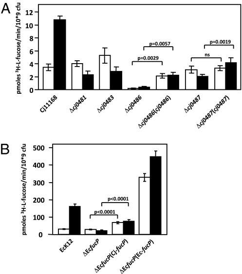
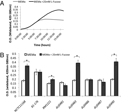
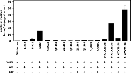
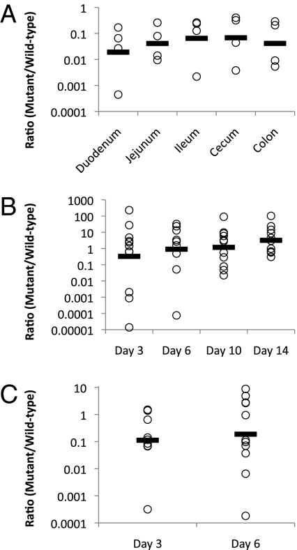
Campylobacter jejuni is a prevalent gastrointestinal pathogen in humans and a common commensal of poultry. When colonizing its hosts, C. jejuni comes into contact with intestinal carbohydrates, including L-fucose, released from mucin glycoproteins. Several strains of C. jejuni possess a genomic island (cj0480c-cj0490) that is up-regulated in the presence of both L-fucose and mucin and allows for the utilization of L-fucose as a substrate for growth. Strains possessing this genomic island show increased growth in the presence of L-fucose and mutation of cj0481, cj0486, and cj0487 results in the loss of the ability to grow on this substrate. Furthermore, mutants in the putative fucose permease (cj0486) are deficient in fucose uptake and demonstrate a competitive disadvantage when colonizing the piglet model of human disease, which is not paralleled in the colonization of poultry. This identifies a previously unrecorded metabolic pathway in select strains of C. jejuni associated with a virulent lifestyle.


Conflict of interest statement

The authors declare no conflict of interest.

Figures

Fig. 1.
Fig. 1.
Complementation of an Escherichia coli fucP mutant. The image shows a MacConkey agar plate supplemented with 30 mM fucose. (A) Positive control, E. coli K12; (B) negative control, E. coli ΔfucP; (C) negative control, E. coli K12+pBR322; (D) positive gene-replacement control, E. coli K12+pBR322+EcfucP; (E and F) replacement with C. jejuni cj0486 in E. coli ΔfucP with pBR322 containing cj0486. Red growth indicates acidification of the medium as a result of fucose fermentation.
Fig. 2.
Fig. 2.
Fucose uptake by C. jejuni (A) and E. coli (B). The initial velocity of [3H]fucose incorporation, which was linear within the first 120 s of the assay, is shown for the indicated strains. White bars: grown in the absence of fucose (uninduced); black bars, grown in the presence of fucose (induced). SDs are displayed by error bars. P values derived from paired t tests compare the statistical differences in uptake rates between the mutants and their complements. ns, no statistically significant difference; i.e., P > 0.05.
Fig. 3.
Fig. 3.
(A) Growth of C. jejuni NCTC 11168 in MEMα medium with and without 25 mM l-fucose over 16 h. (B) Maximum OD attained over 36 h of growth for the reference strains C. jejuni NCTC 11168, 81–176, and RM1221, and the mutant strains C. jejuni Δcj0481, Δcj0483, Δcj0486, Δcj0487, and Δcj0490. The asterisk denotes statistical significance, P < 0.05 using a paired t test. Error bars represent the SD.
Fig. 4.
Fig. 4.
ATP-dependent 3H-l-fucose phosphorylation and GTP-dependent 3H-l-fucose activation. Whole-cell lysates (100 μg) of the indicated strains grown in the presence or absence of [3H]-l-fucose (as indicated) for 24 h and assayed for their potential to fucose under the indicated assay conditions. Ec, E. coli K12 wild-type; ΔEcfucK, E. coli K12 fucK mutant; Cj, C. jejuni NCTC11168 wild-type; Δcj0486, C. jejuni NCTC11168 Δ0486; Bt, B. thetaiotaomicron wild-type. The presence or absence of ATP and GTP are indicated by (+) and (−). Formation of modified 3H-l-fucose was found to be linear over the first 10 min of the assay. SDs are displayed by error bars. 3H-l-fucose alone did not show any significant binding to the column material.
Fig. 5.
Fig. 5.
(A) Competitive C. jejuni colonization assay in neonatal piglets. The points represent the ratio of Δcj0486 mutant/wild-type NCTC 11168 recovered from each intestinal segment indicated, 3 d postinoculation. Values are adjusted relative to the ratio of the inoculum of 0.927. A line denotes the mean for each dataset where n = 4. Statistical significance was determined by a one-way ANOVA, P < 0.002. (B) Competitive C. jejuni colonization assay of chicks over a 2-wk period postinoculation. Each point is the ratio of mutant to wild-type recovered from the cecum of a single chick. A line denotes the mean for each day, n = 9–11 per day. Statistical significance was determined by a one-way ANOVA, P = 0.82. (C) Competitive C. jejuni colonization assay in chicks receiving an l-fucose dietary supplement over a 6-d period postinoculum. Day 3, n = 9 and day 6, n = 11. Statistical significance was determined by a Student t test for each day.

Similar articles

Cited by

References

    1. Coker AO, Isokpehi RD, Thomas BN, Amisu KO, Obi CL. Human campylobacteriosis in developing countries. Emerg Infect Dis. 2002;8:237–244. - PMC - PubMed
    1. Galanis E. Campylobacter and bacterial gastroenteritis. CMAJ. 2007;177:570–571. - PMC - PubMed
    1. Ketley JM. Pathogenesis of enteric infection by Campylobacter. Microbiology. 1997;143(1):5–21. - PubMed
    1. Lee A, O'Rourke JL, Barrington PJ, Trust TJ. Mucus colonization as a determinant of pathogenicity in intestinal infection by Campylobacter jejuni: A mouse cecal model. Infect Immun. 1986;51:536–546. - PMC - PubMed
    1. Byrne CM, Clyne M, Bourke B. Campylobacter jejuni adhere to and invade chicken intestinal epithelial cells in vitro. Microbiology. 2007;153:561–569. - PubMed

Publication types