. 2011 Apr 26;108(17):7046-51.
doi: 10.1073/pnas.1018380108. Epub 2011 Apr 11.

Orthograde dihydropyridine receptor signal regulates ryanodine receptor passive leak

Affiliations

Orthograde dihydropyridine receptor signal regulates ryanodine receptor passive leak

José Miguel Eltit et al. Proc Natl Acad Sci U S A. .

Abstract

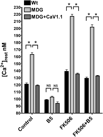
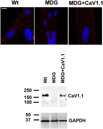
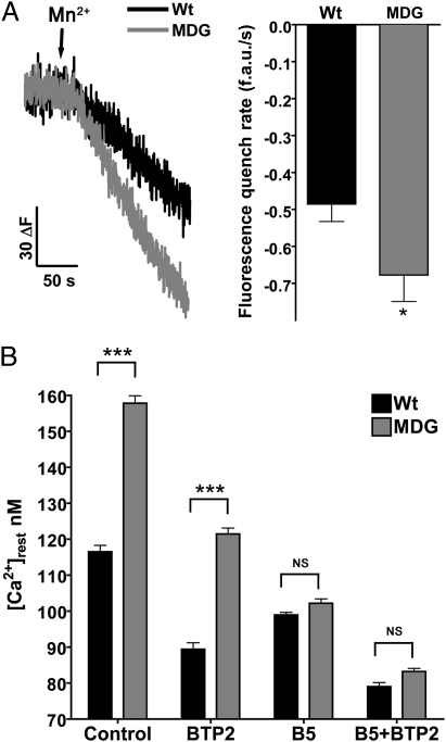
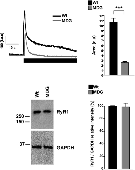
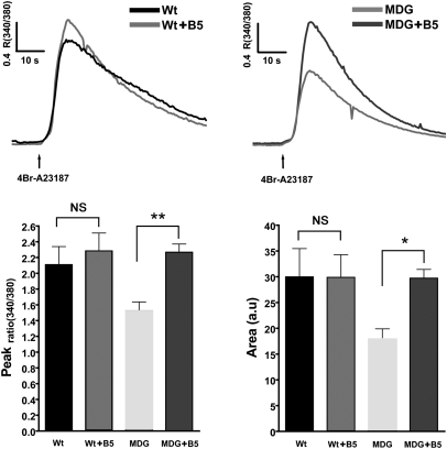
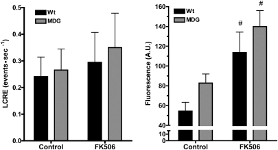
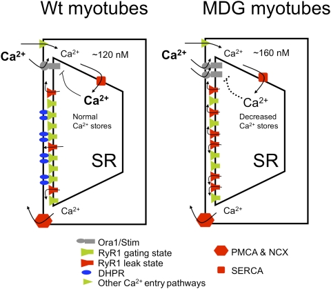
The skeletal muscle dihydropyridine receptor (DHPR) and ryanodine receptor (RyR1) are known to engage a form of conformation coupling essential for muscle contraction in response to depolarization, referred to as excitation-contraction coupling. Here we use WT and Ca(V)1.1 null (dysgenic) myotubes to provide evidence for an unexplored RyR1-DHPR interaction that regulates the transition of the RyR1 between gating and leak states. Using double-barreled Ca(2+)-selective microelectrodes, we demonstrate that the lack of Ca(V)1.1 expression was associated with an increased myoplasmic resting [Ca(2+)] ([Ca(2+)](rest)), increased resting sarcolemmal Ca(2+) entry, and decreased sarcoplasmic reticulum (SR) Ca(2+) loading. Pharmacological control of the RyR1 leak state, using bastadin 5, reverted the three parameters to WT levels. The fact that Ca(2+) sparks are not more frequent in dysgenic than in WT myotubes adds support to the hypothesis that the leak state is a conformation distinct from gating RyR1s. We conclude from these data that this orthograde DHPR-to-RyR1 signal inhibits the transition of gated RyR1s into the leak state. Further, it suggests that the DHPR-uncoupled RyR1 population in WT muscle has a higher propensity to be in the leak conformation. RyR1 leak functions are to keep [Ca(2+)](rest) and the SR Ca(2+) content in the physiological range and thus maintain normal intracellular Ca(2+) homeostasis.


Conflict of interest statement

The authors declare no conflict of interest.

Figures

Fig. 1.
Fig. 1.
B5 equalizes [Ca2+]rest among CaV1.1-expressing and dysgenic (MDG) myotubes. [Ca2+]rest determinations were done in WT (n = 18), dysgenic (MDG, n = 17), and dysgenic myotubes transduced with CaV1.1 lentivirus (MDG+CaV1.1, n = 16). Where indicated, the measurements were done in the presence of 20 μM B5 (WT n = 18, dysgenic n = 16, and dysgenic+CaV1.1 n = 10), FK506 (WT n = 10, dysgenic n = 10, and dysgenic+CaV1.1 n = 10), or FK506 plus B5 (FK506+B5, WT n = 10, dysgenic n = 10, and dysgenic+CaV1.1 n = 10). Mean ± SEM is shown. *P < 0.001 (ANOVA). NS, not significant.
Fig. 2.
Fig. 2.
Dysgenic (MDG) myotubes do not express the α1S subunit of the DHPR (CaV1.1). The expression of CaV1.1 was assessed by immunostaining in WT, dysgenic (MDG), and dysgenic (MDG)+CaV1.1 myotubes. Clusters of DHPRs (red foci) are located at the cell surface in WT and in dysgenic+CaV1.1 but not in dysgenic myotubes. Western blot confirms lack of CaV1.1 expression in dysgenic myotubes and its rescue by stable transduction. (Scale bar, 10 μm.)
Fig. 3.
Fig. 3.
Resting Ca2+ entry is increased in dysgenic myotubes. Resting Ca2+ entry was estimated using the Mn+2 quench technique. (A) Representative traces for WT and dysgenic are shown. The fluorescence decay induced by Mn2+ permeability was fitted to a linear regression for each response: WT (n = 32) and dysgenic (MDG) (n = 25). Mean ± SEM is shown. *P < 0.05 (t test). (B) [Ca2+]rest was measured as described in Materials and Methods. The measurements were done in WT (n = 12) and dysgenic (MDG) myotubes (n = 11) in control conditions, treated with 5 μM BTP2 alone (WT n = 10, dysgenic n = 11), B5 alone (WT n = 17, dysgenic n = 18), or the combination of both drugs (B5+BTP2, WT n = 10, dysgenic n = 10). Results are shown as mean ± SEM. ***P < 0.001 (ANOVA). NS, not significant.
Fig. 4.
Fig. 4.
Dysgenic myotubes have decreased releasable SR Ca2+. WT and dysgenic myotubes loaded with Fluo-5N were exposed to 20 mM caffeine in free Ca2+ media (1 mM EGTA) as describes in Materials and Methods. Representative traces are shown for WT and dysgenic (MDG) myotubes; the black rectangle shows when caffeine was applied. The area under the curve of each response was calculated [WT n = 66 and dysgenic (MDG) n = 69], and the data are shown as mean ± SEM. ***P < 0.001 (ANOVA) (Upper). Western blot analysis shows that RyR1 expression is not different between WT and dysgenic myotubes [Lower; WT n = 3 and dysgenic (MDG) n = 3].
Fig. 5.
Fig. 5.
Intracellular Ca2+ content is decreased in dysgenic myotubes and is corrected by B5. WT and dysgenic myotubes were loaded with the ratiometric dye Fura-4F. The total Ca2+ released by 4Br-A23187 in Ca2+-free solution was measured as an estimation of the Ca2+SR loading of the myotubes. Mean traces of the responses [WT black trace and dysgenic (MDG) trace] and the effect of 20 μM B5 are shown [WT+B5 trace and dysgenic (MDG)+B5 trace]. Lower: Graphs show peak and area values plotted as mean ± SEM [WT n = 20, dysgenic (MDG) n = 29, WT+B5 n = 24, dysgenic (MDG)+B5 n = 15]. *P < 0.05; **P < 0.01 (ANOVA).
Fig. 6.
Fig. 6.
Increased [Ca2+]rest in dysgenic myotubes does not correlate with frequency of spontaneous Ca2+sparks. Spontaneous LCREs were measured as described in Materials and Methods on WT and dysgenic (MDG) myotubes. The frequency of spontaneous LCRE was calculated for each individual cell, and the data are shown as mean ± SEM (Left; WT n = 10, dysgenic n = 10). In addition, the global fluorescence of each myotube was calculated and plotted (Right; WT n = 10, dysgenic n = 10) as mean ± SEM. #P < 0.05 (ANOVA). The effect of FK506 treatment on both LCRE frequency and global fluorescence is shown.
Fig. 7.
Fig. 7.
Altered resting Ca2+ homeostasis in dysgenic (MDG) myotubes (a model). We propose a model in which association with the DHPR does not allow RyRs to enter into the leak state. This limits the availability of leaky channels and preserves a restricted passive Ca+2 efflux from the SR. At equilibrium the SR Ca2+ content is not depleted, and negative feedback controls the plasmalemmal Ca+2 permeability (Left). In the absence of DHPRs, in dysgenic (MDG) myotubes, there is an increased passive Ca2+ efflux from the SR through RyR1, resulting in an increase of [Ca2+]rest and a decrease of Ca2+content in the SR, thus activating store operated calcium entry. The overall effect is an increase of the [Ca2+]rest (Right). SERCA, sarco-endoplasmic Ca2+ ATPase; PMCA, plasma membrane Ca2+ ATPase; NCX, Ca2+/Na+ exchanger of the plasma membrane.

Similar articles

Cited by

References

    1. Perez CF, López JR, Allen PD. Expression levels of RyR1 and RyR3 control resting free Ca2+ in skeletal muscle. Am J Physiol Cell Physiol. 2005;288:C640–C649. - PubMed
    1. Berridge MJ, Bootman MD, Roderick HL. Calcium signalling: Dynamics, homeostasis and remodelling. Nat Rev Mol Cell Biol. 2003;4:517–529. - PubMed
    1. Turner PR, Westwood T, Regen CM, Steinhardt RA. Increased protein degradation results from elevated free calcium levels found in muscle from mdx mice. Nature. 1988;335:735–738. - PubMed
    1. Yang T, et al. Elevated resting [Ca(2+)](i) in myotubes expressing malignant hyperthermia RyR1 cDNAs is partially restored by modulation of passive calcium leak from the SR. Am J Physiol Cell Physiol. 2007;292:C1591–C1598. - PubMed
    1. Lopez JR, Gerardi A, Lopez MJ, Allen PD. Effects of dantrolene on myoplasmic free [Ca2+] measured in vivo in patients susceptible to malignant hyperthermia. Anesthesiology. 1992;76:711–719. - PubMed

Publication types

MeSH terms