. 2011 Apr 26;108(17):7166-71.
doi: 10.1073/pnas.1016086108. Epub 2011 Apr 11.

HDL promotes rapid atherosclerosis regression in mice and alters inflammatory properties of plaque monocyte-derived cells

Affiliations

HDL promotes rapid atherosclerosis regression in mice and alters inflammatory properties of plaque monocyte-derived cells

Jonathan E Feig et al. Proc Natl Acad Sci U S A. .

Erratum in

  • Proc Natl Acad Sci U S A. 2011 Aug 23;108(34):14371

Abstract

HDL cholesterol (HDL-C) plasma levels are inversely related to cardiovascular disease risk. Previous studies have shown in animals and humans that HDL promotes regression of atherosclerosis. We hypothesized that this was related to an ability to promote the loss of monocyte-derived cells (CD68(+), primarily macrophages and macrophage foam cells) from plaques. To test this hypothesis, we used an established model of atherosclerosis regression in which plaque-bearing aortic arches from apolipoprotein E-deficient (apoE(-/-)) mice (low HDL-C, high non-HDL-C) were transplanted into recipient mice with differing levels of HDL-C and non-HDL-C: C57BL6 mice (normal HDL-C, low non-HDL-C), apoAI(-/-) mice (low HDL-C, low non-HDL-C), or apoE(-/-) mice transgenic for human apoAI (hAI/apoE(-/-); normal HDL-C, high non-HDL-C). Remarkably, despite persistent elevated non-HDL-C in hAI/apoE(-/-) recipients, plaque CD68(+) cell content decreased by >50% by 1 wk after transplantation, whereas there was little change in apoAI(-/-) recipient mice despite hypolipidemia. The decreased content of plaque CD68(+) cells after HDL-C normalization was associated with their emigration and induction of their chemokine receptor CCR7. Furthermore, in CD68(+) cells laser-captured from the plaques, normalization of HDL-C led to decreased expression of inflammatory factors and enrichment of markers of the M2 (tissue repair) macrophage state. Again, none of these beneficial changes were observed in the apoAI(-/-) recipients, suggesting a major requirement for reverse cholesterol transport for the beneficial effects of HDL. Overall, these results establish HDL as a regulator in vivo of the migratory and inflammatory properties of monocyte-derived cells in mouse atherosclerotic plaques, and highlight the phenotypic plasticity of these cells.


Conflict of interest statement

The authors declare no conflict of interest.

Figures

Fig. 1.
Fig. 1.
ApoAI deficiency impairs plaque regression despite hypolipidemia, but selective normalization of plasma HDL-C levels promotes plaque regression despite hyperlipidemia. (A) After 16 wk on the Western diet, apoE−/− mice were killed and aortic arches were used for baseline determinations (Baseline), or were transplanted into WT or apoAI−/− recipients. Plaque size and the area immunostained by an anti-CD68 antibody were determined. *P < 0.05, compared with Baseline or the apoAI recipients. (B) Similar to A, but transplantation was into apoE−/−, or apoE−/− mice expressing the human apoAI transgene (humanApoAI/apoE−/−). *P < 0.05, compared with Baseline or apoE−/− recipients. For all figures, the number of mice were: Baseline, n = 10; recipients: WT, n = 11; apoAI−/−, n = 14; apoE−/−, n = 11; hAI/apoE−/−, n = 13.
Fig. 2.
Fig. 2.
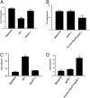
Effects of HDL-C and apoAI deficiency on the ability of Ly-6Chi -derived cells to emigrate from plaques and on the induction of chemokine receptor CCR7 in plaque CD68+ cells. (A and B) One week before transplantation, circulating Ly-6Chi monocytes in donor (apoE−/−) mice were labeled with fluorescent beads (22, 23). One week after transplantation, aortic arches were harvested and sectioned. The number of beads in the plaque sections was counted using fluorescence microscopy. *P < 0.05, compared with Baseline. (C and D) CD68+ cells were laser-captured from the plaques from donor mice (Baseline) and recipient mice. CCR7 levels were then measured in the isolated RNA by qRT-PCR. For A and B, the asterisk corresponds to statistical significance, P < 0.05, compared with Baseline or apoAI−/− recipient mice. For C and D, the asterisk corresponds to statistical significance, P < 0.05, compared with Baseline or apoE−/− recipient mice.
Fig. 3.
Fig. 3.
CCR7 gene expression is regulated in vitro by cholesterol loading and efflux. CCR7 gene expression (normalized to cyclophilin A) was measured in a model of immature dendritic cells, DC2.4, under the following conditions: basal, after 24 h of incubation with acetylated LDL (AcLDL), after 24 h of incubation with HDL3 to promote cholesterol efflux under basal conditions, or after 24 h of loading with acetylated LDL, followed by incubation for 24 h with HDL3 (Load/Unload). Shown are the means ± SEM from a representative experiment performed in triplicate. *P < 0.01 vs. basal state.
Fig. 4.
Fig. 4.
Normalization of HDL-C levels change the gene expression of markers of inflammation and of the M2 macrophage phenotype in plaque CD68+ cells in an apoAI-dependent manner. CD68+ cells were laser captured from the plaques of donor (Baseline) mice or from grafts 1 wk after transplantation into recipients. The mRNA levels of the indicated genes associated with inflammation (A) or the M2 macrophage state (B) were measured by qRT-PCR. Data are based on two pools of RNA, each consisting of three independent mice, and are expressed as mean (± SEM) fold-change over Baseline. ICAM-1, intercellular adhesion molecule 1; MR, mannose receptor. *P < 0.05 vs. Baseline.
Fig. 5.
Fig. 5.
HDL and apoAI induce in vitro the gene expression of markers of M2 macrophages. BMDMs were isolated from WT mice, incubated for 6 d in the presence of M-CSF. HDL3 or apoAI was added for 5 h and gene expression of arginase 1, Fizz 1, and cycophilin A were assessed by qRT-PCR. Shown are the results (mean +/− SEM, normalized to cyclophilin A) of three experiments, each done in duplicate or triplicate. *P < 0.01, **P < 0.001 vs. corresponding control value.
Fig. 6.
Fig. 6.
Effects of the plasma lipoprotein environment on the protein expression of arginase I in plaques. Aortic sections from mice in each group described in Fig. 4 were immunostained for arginase I using a fluorescently tagged secondary antibody. Representative laser-confocal microscopic images are displayed (40×); L, lumen. The wavy lines are autofluorescent internal elastic lamina.

References

    1. Castelli WP, et al. Incidence of coronary heart disease and lipoprotein cholesterol levels. The Framingham Study. JAMA. 1986;256:2835–2838. - PubMed
    1. Feig JE, Shamir R, Fisher EA. Atheroprotective effects of HDL: Beyond reverse cholesterol transport. Curr Drug Targets. 2008;9:196–203. - PubMed
    1. Franceschini G. Epidemiologic evidence for high-density lipoprotein cholesterol as a risk factor for coronary artery disease. Am J Cardiol. 2001;88(12A):9N–13N. - PubMed
    1. Gordon DJ, Rifkind BM. High-density lipoprotein—The clinical implications of recent studies. N Engl J Med. 1989;321:1311–1316. - PubMed
    1. Miller NE, Miller GJ. Letter: High-density lipoprotein and atherosclerosis. Lancet. 1975;1:1033. - PubMed

Publication types

MeSH terms