. 2011 Apr 26;108(17):7177-82.
doi: 10.1073/pnas.1103350108. Epub 2011 Apr 11.

Targeting p21-activated kinase 1 (PAK1) to induce apoptosis of tumor cells

Affiliations

Targeting p21-activated kinase 1 (PAK1) to induce apoptosis of tumor cells

Christy C Ong et al. Proc Natl Acad Sci U S A. .

Abstract

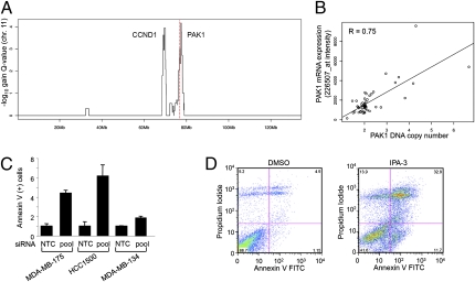
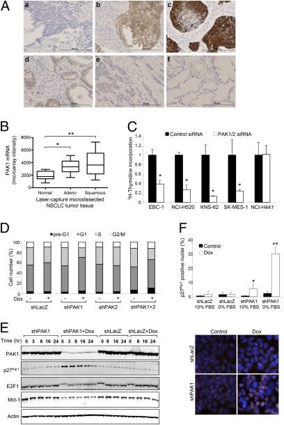
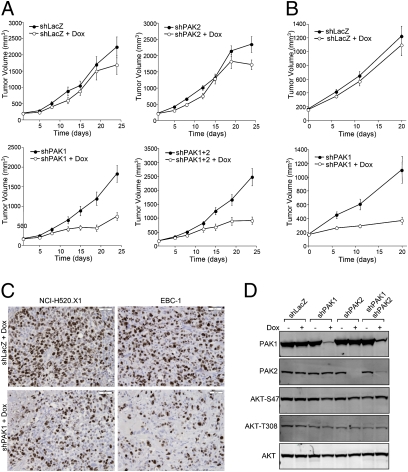
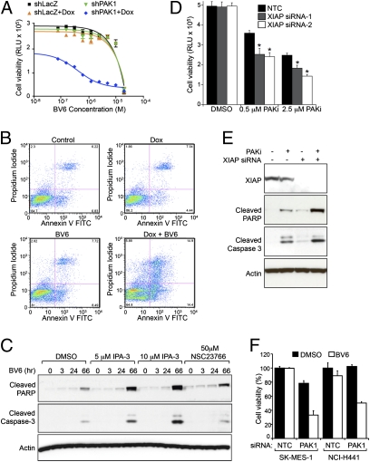
p21-activated kinases (PAKs) are serine/threonine protein kinases that serve as important mediators of Rac and Cdc42 GTPase function as well as pathways required for Ras-driven tumorigenesis. PAK1 has been implicated in signaling by growth factor receptors and morphogenetic processes that control cell polarity, invasion, and actin cytoskeleton organization. To better understand the role of PAK1 in tumorigenesis, PAK1 genomic copy number and expression were determined for a large panel of breast, lung, and head and neck tumors. PAK1 genomic amplification at 11q13 was prevalent in luminal breast cancer, and PAK1 protein expression was associated with lymph node metastasis. Breast cancer cells with PAK1 genomic amplification rapidly underwent apoptosis after inhibition of this kinase. Strong nuclear and cytoplasmic PAK1 expression was also prevalent in squamous nonsmall cell lung carcinomas (NSCLCs), and selective PAK1 inhibition was associated with delayed cell-cycle progression in vitro and in vivo. NSCLC cells were profiled using a library of pathway-targeted small-molecule inhibitors, and several synergistic combination therapies, including combination with antagonists of inhibitor of apoptosis proteins, were revealed for PAK1. Dual inhibition of PAK1 and X chromosome-linked inhibitor of apoptosis efficiently increased effector caspase activation and apoptosis of NSCLC cells. Together, our results provide evidence for dysregulation of PAK1 in breast and squamous NSCLCs and a role for PAK1 in cellular survival and proliferation in these indications.


Conflict of interest statement

Conflict of interest statement: C.C.O., P.M.H., W.Z., V.T., T.T., T.O., D.V., M.B., L.S.F., E.M.B., H.K., and K.P.H. are employees of Genentech, Inc.

Figures

Fig. 1.
Fig. 1.
Analysis of PAK1 genomic amplification and functional role in human breast tumors. (A) GISTIC analysis of 11q13 copy-number gains. The red line represents the location of the PAK1 gene. (B) PAK1 DNA copy and mRNA expression are associated for 51 breast tumor samples (Pearson correlation = 0.75). (C) Increased Annexin V-positive cells after knockdown of PAK1 expression for three breast cancer cell lines with focal PAK1 genomic amplification and estrogen receptor-positive status: MDA-MB-175, HCC1500, and MDA-MB-134 IV. (D) FACS analysis for MDA-MB-175 cells treated with IPA-3 for 48 h. Annexin V/PI staining was then done to assess apoptosis/necrosis.
Fig. 2.
Fig. 2.
PAK1 is highly expressed in human lung tumors and plays a critical role in proliferation of squamous NSCLC cell lines. (A) IHC for PAK1 was performed on (A, ac) squamous NSCLC, (A, d and e) adenocarcinoma NSCLC, and (A, f) normal lung tissues. (Scale bar, 200 μm.) (B) PAK1 mRNA expression in laser-capture microdissected lung tissues. Relative to normal tissues (n = 9), PAK1 expression was greater in squamous (n = 16; **P = 0.0005) and adenocarcinoma NSCLC (n = 29; *P = 0.008). (C) Proliferation of squamous NSCLC cell lines transfected with siRNA oligonucleotides was measured by [3H]thymidine uptake assay. The data are normalized to control and shown as the mean ± SD. (D) Accumulation of cells in G1 phase of the cell cycle is evident after inducible PAK1 knockdown in NCI-H520.X1 cells. (E) NCI-H520.X1 cells were serum-starved for 24 h, and cell-cycle reentry was monitored by harvesting cell lysates at the indicated time points after growth in 10% serum-containing media. (F) Anti-p27Kip1 (red) and DAPI (blue) immunofluorescence after PAK1-inducible knockdown. Quantification of p27Kip1 nuclear accumulation (n = 2,000 cells). *P < 0.05. **P < 0.0001.
Fig. 3.
Fig. 3.
PAK1 is required for growth of established NCI-H520.X1 and EBC-1 squamous NSCLC tumors. (A) NCI-H520.X1 cells expressing inducible shRNAs against LacZ, PAK1, PAK2, or PAK1+PAK2 were implanted in the flank of athymic mice. Doxycycline was administered when tumors were established and resulted in inhibition of tumor growth for mice bearing shPAK1 and shPAK1+2 NCI-H520.X1 cells. (B) EBC-1 shLacZ or shPAK1 tumor-bearing mice were administered doxycycline to inhibit PAK1. (C) Histologic analysis of in vivo knockdown of PAK1 in NCI-H520.X1 and EBC-1 tumors. (D) AKT phosphorylation was not altered on PAK1 and/or PAK2 knockdown in squamous NSCLC tumors.
Fig. 4.
Fig. 4.
PAK1 inhibition combines with IAP antagonists to promote apoptosis of NSCLC cells. (A) EBC-1-shPAK1 and -shLacZ cells were treated with Dox and BV6 IAP antagonist. The highest concentration of BV6 was 20 μM, and twofold serial dilutions were assessed in quadruplicate. (B) FACS analysis for Annexin V and PI staining to assess apoptosis/necrosis after PAK1 and IAP antagonism. (C) Combinatorial accumulation of cleaved PARP and caspase-3 resulting from IAP antagonism with IPA-3 or NSC23766. (D) Down-regulation of XIAP expression potentiates the proapoptotic activity of PF-3758309 PAK small-molecule inhibitor (PAK SMI; P < 0.0001). Cells were transfected with siRNA oligonucleotides for 48 h before inhibitor treatment for an additional 72 h. (E) Combined antagonism of XIAP and PAK1 promotes efficient cleavage of PARP and caspase-3. (F) Dual PAK1 and IAP inhibition results in a synergistic decrease in viability of SK-Mes-1 (squamous) and NCI-H441 (adenocarcinoma) NSCLC cells (P < 0.0001).

References

    1. Arias-Romero LE, Chernoff J. A tale of two Paks. Biol Cell. 2008;100:97–108. - PubMed
    1. Sells MA, et al. Human p21-activated kinase (Pak1) regulates actin organization in mammalian cells. Curr Biol. 1997;7:202–210. - PubMed
    1. Delorme V, et al. Cofilin activity downstream of Pak1 regulates cell protrusion efficiency by organizing lamellipodium and lamella actin networks. Dev Cell. 2007;13:646–662. - PMC - PubMed
    1. Balasenthil S, et al. p21-activated kinase-1 signaling mediates cyclin D1 expression in mammary epithelial and cancer cells. J Biol Chem. 2004;279:1422–1428. - PubMed
    1. Schraml P, et al. Combined array comparative genomic hybridization and tissue microarray analysis suggest PAK1 at 11q13.5-q14 as a critical oncogene target in ovarian carcinoma. Am J Pathol. 2003;163:985–992. - PMC - PubMed

Publication types

MeSH terms