. 2011 Apr 26;108(17):6957-62.
doi: 10.1073/pnas.1018777108. Epub 2011 Apr 11.

Mice lacking phosphatase PP2A subunit PR61/B'delta (Ppp2r5d) develop spatially restricted tauopathy by deregulation of CDK5 and GSK3beta

Affiliations

Mice lacking phosphatase PP2A subunit PR61/B'delta (Ppp2r5d) develop spatially restricted tauopathy by deregulation of CDK5 and GSK3beta

Justin V Louis et al. Proc Natl Acad Sci U S A. .

Abstract

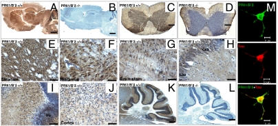
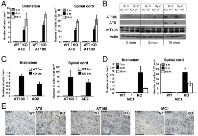
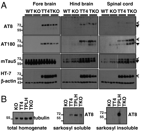
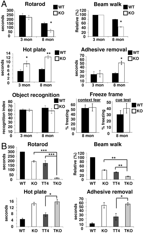
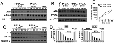
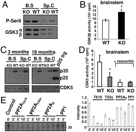
Functional diversity of protein phosphatase 2A (PP2A) enzymes mainly results from their association with distinct regulatory subunits. To analyze the functions of one such holoenzyme in vivo, we generated mice lacking PR61/B'δ (B56δ), a subunit highly expressed in neural tissues. In PR61/B'δ-null mice the microtubule-associated protein tau becomes progressively phosphorylated at pathological epitopes in restricted brain areas, with marked immunoreactivity for the misfolded MC1-conformation but without neurofibrillary tangle formation. Behavioral tests indicated impaired sensorimotor but normal cognitive functions. These phenotypical characteristics were further underscored in PR61/B'δ-null mice mildly overexpressing human tau. PR61/B'δ-containing PP2A (PP2A(T61δ)) poorly dephosphorylates tau in vitro, arguing against a direct dephosphorylation defect. Rather, the activity of glycogen synthase kinase-3β, a major tau kinase, was found increased, with decreased phosphorylation of Ser-9, a putative cyclin-dependent kinase 5 (CDK5) target. Accordingly, CDK5 activity is decreased, and its cellular activator p35, strikingly absent in the affected brain areas. As opposed to tau, p35 is an excellent PP2A(T61δ) substrate. Our data imply a nonredundant function for PR61/B'δ in phospho-tau homeostasis via an unexpected spatially restricted mechanism preventing p35 hyperphosphorylation and its subsequent degradation.


Conflict of interest statement

The authors declare no conflict of interest.

Figures

Fig. 1.
Fig. 1.
PR61/B’δ localization in brain, spinal cord, and PC-12 cells. Immunohistochemistry staining (3-step procedure) of PR61/B’δ in WT brain (A) and spinal cord (C). Brain (B) and spinal cord (D) of PR61/B’δ KO mice are shown as control. Detailed expression of PR61/B’δ in WT striatum (E), thalamus (F), brain stem (G), CA1, 2 and 3 regions (H) and dentate gyrus (DG) of hippocampus (I), cortex (J), and cerebellum (K). KO cerebellum is shown as control (L). Nuclei are counterstained with haematoxylin (blue). Scale bars: 1 mm (AD, K, and L); 50 μm (EJ). (M) Immunofluorescence showing colocalization of PR61/B’δ (green) and tau (red) in NGF-differentiated PC12 cells (Scale bar: 10 μm).
Fig. 2.
Fig. 2.
Age-related tau hyperphosphorylation and misfolding in brain stem and spinal cord of PR61/B’δ KO mice. (A) Immunohistochemistry (IHC) with AT8 (Ser202/Thr205) and AT180 (Thr231) showing progressive tau phosphorylation in brain stem (left) and dorsal horn of the cervical spinal cord (right) of PR61/B’δ KO mice. (B) Western blot of brain stem and spinal cord lysates (n = 5) developed with the indicated antibodies. (C) IHC with AT100 (Ser212/Ser214) and AD2 (Ser396/Ser404) at six months in brainstem (left) and spinal cord (right). (D) IHC with MC1 (recognizing misfolded tau) in KO and age-matched WT brain stem (left) and spinal cord (right). Only cytoplasmic tau stainings were quantified (A, C, and D) with Leica QWin V2.8 software. (E) Representative IHC images from brainstem at six months (for complete overview, see Fig. S4). Scale bars: 100 μM.
Fig. 3.
Fig. 3.
Tau phosphorylation in TKO mice. (A) Forebrain, hindbrain (brain stem/cerebellum), and spinal cord extracts of three independent WT, KO, TT4, and TKO mice (10 months) subjected to Western blotting with the indicated antibodies. Open arrowheads: transgenic human tau; filled arrowheads: endogenous tau. (B) Western blots with the indicated antibodies of total brain lysates and sarkosyl (in)soluble fractions of these lysates from KO, TT4, TKO, and TPLH mice.
Fig. 4.
Fig. 4.
Behavior tests in age- and gender-matched WT, KO, TT4, and TKO mice. (A) In three- or eight-month-old PR61/B’δ KO mice compared to WT (n = 6 for each condition). (B) In 10-month-old TKO mice compared to TT4, KO, and WT (n = 6 for each condition). Statistical significance was evaluated with Student’s t-test (* : p < 0.05; ** : p < 0.01; *** : p < 0.001).
Fig. 5.
Fig. 5.
In vitro tau dephosphorylation with various PP2A holoenzymes. (A) Equilibrated amounts of de novo purified PP2AD and PP2AT55 were incubated with phospho-tau isolated from TPLH mice for the indicated times. Dephosphorylation was monitored with AT8 and AT180 antibodies. (B) Same experiment with different amounts of PP2AD (units indicated). (C) Same experiment with PP2AT55α and PP2AT61δ isolated from GST-PR55α and GST-PR61δ expressing COS7 cells. Normalized quantifications in D. (E) [32P]-tau, in vitro labeled by CDK2/cyclinA was incubated with the indicated amounts of PP2AT61δ or PP2AT55α. At several time points aliquots were taken and TCA soluble [32P] measured.
Fig. 6.
Fig. 6.
GSK3β and CDK5 status in PR61/B’δ KO brainstem and spinal cord. (A) Western blot of brain stem and spinal cord extracts of 18-month-old mice (n = 5) showing decreased GSK3β Ser9 phosphorylation in KO mice. (B) Increased activity in GSK3β IPs from brain stem extracts of 18-month-old KO mice (n = 5) measured with phospho-glycogen synthase peptide 2 substrate. (C) Western blot showing highly reduced levels of p35 in brain stem and spinal cord of three- and 18-month-old KO mice. Positive control: brain extract of p25 transgenic (trg) mice (42). (D) Decreased activity in CDK5 IPs from brain stem extracts of six-month-old KO mice (n = 5) measured with Histone IIA as substrate and roscovitin (10 μM) as specificity control. (E) [32P]-p35, in vitro labeled by CDK2/cyclinA was incubated for the indicated times with PP2AT61δ, PP2AT55α, PP2AD, or PP1 (all 1 U/ml). Dephosphorylation was monitored by autoradiography following SDS/PAGE and quantified (averages of two independent experiments).

References

    1. Janssens V, Longin S, Goris J. PP2A holoenzyme assembly: In cauda venenum (the sting is in the tail) Trends Biochem Sci. 2008;33:113–121. - PubMed
    1. Eichhorn PJ, Creyghton MP, Bernards R. Protein phosphatase 2A regulatory subunits and cancer. Biochim Biophys Acta. 2009;1795:1–15. - PubMed
    1. Janssens V, Goris J, Van Hoof C. PP2A: The expected tumor suppressor. Curr Opin Genet Dev. 2005;15:34–41. - PubMed
    1. Tian Q, Wang J. Role of serine/threonine protein phosphatase in Alzheimer’s disease. Neurosignals. 2002;11:262–269. - PubMed
    1. Götz J, Probst A, Ehler E, Hemmings B, Kues W. Delayed embryonic lethality in mice lacking protein phosphatase 2A catalytic subunit Calpha. Proc Natl Acad Sci USA. 1998;95:12370–12375. - PMC - PubMed

Publication types

MeSH terms