. 2011 Jun 3;286(22):19576-88.
doi: 10.1074/jbc.M110.196311. Epub 2011 Apr 10.

Daxx-beta and Daxx-gamma, two novel splice variants of the transcriptional co-repressor Daxx

Affiliations

Daxx-beta and Daxx-gamma, two novel splice variants of the transcriptional co-repressor Daxx

Nils Wethkamp et al. J Biol Chem. .

Abstract

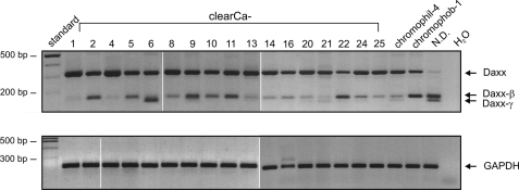
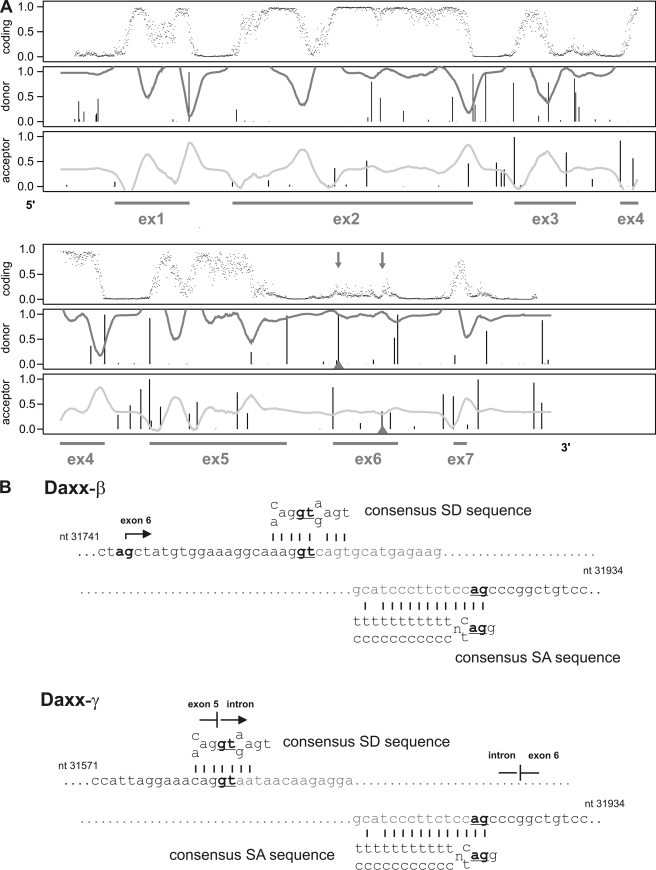
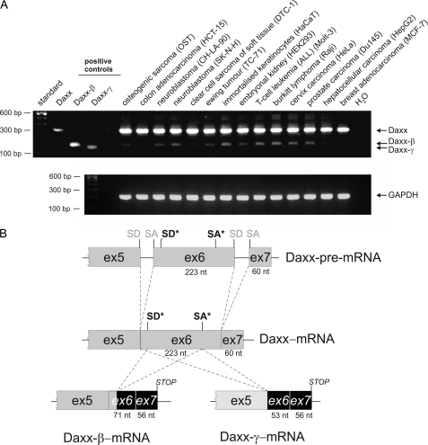
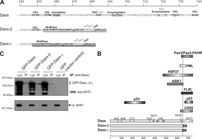
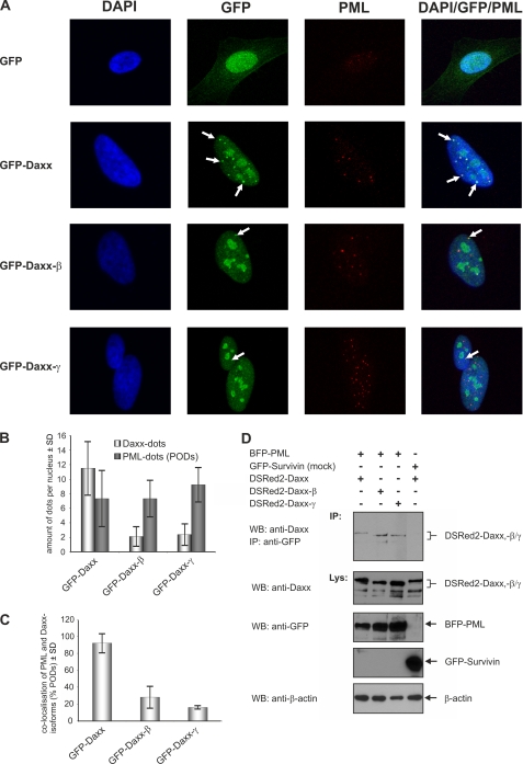
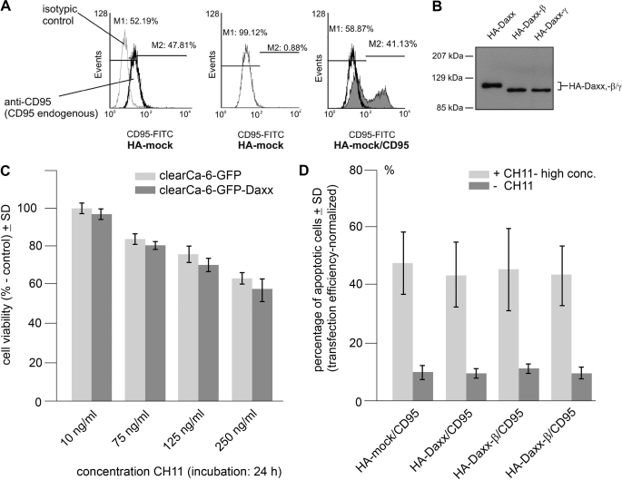
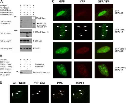
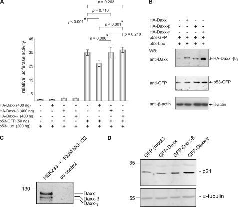
Daxx is involved in transcriptional control and apoptosis. It comprises several domains, including a regulatory C terminus that is responsible for the interaction with numerous proteins such as p53, promyelocytic leukemia protein (PML), and Hsp27. Here, we describe the identification and characterization of two novel variants of Daxx termed Daxx-β and Daxx-γ, which are generated by alternative splicing. Alternative splicing results in a truncated regulatory C terminus in both proteins. As a consequence, Daxx-β and Daxx-γ show a markedly decreased affinity to PML, which in turn is associated with a different subnuclear localization of these proteins compared with Daxx. Although Daxx is localized mainly in PML-oncogenic domains (PODs) Daxx-β and Daxx-γ display a distinct distribution pattern. Furthermore, Daxx-β and Daxx-γ show a decreased affinity to p53 also due to the truncated C terminus. We provide evidence that the p53 recruitment into PODs is Daxx isoform-dependent. The decreased affinity of Daxx-β/-γ to p53 and PML results in a diffuse localization of p53 throughout the nucleus. In contrast to Daxx, Daxx-β and Daxx-γ are unable to repress p53-mediated transcription. Therefore, alternative splicing of Daxx might indicate an additional level in the cellular apoptosis network.


Figures

FIGURE 1.
FIGURE 1.
Expression of different Daxx mRNA variants in various RCC cell lines of all major histological types. Besides the expected 316-bp PCR amplification product two additional smaller fragments were generated. GAPDH PCRs were performed as control. Abbreviations: clearCa, clear cell renal carcinomas; chromophil, papillary/chromophilic renal cell carcinoma; chromophob, chromophobic renal cell carcinoma; N.D., the histological subtype of this RCC line is not determined.
FIGURE 2.
FIGURE 2.
Detection of potential alternative splice donor (SD) and splice acceptor (SA) sites in exon 6 of the Daxx gene. A, NetGene2 analysis of the genomic Daxx sequence. The upper panel of the output predicts the coding region whereas values close to 0 indicate an intron region, and values close to 1 indicate exons. Prediction of SD and SA is given below with a confidence level of 90% (30, 31). The underlying exon progression of the Daxx gene is marked by the string (GenBank accession no. Z97183). B, comparison of the alternative SA and SD sites of exon 6 with the consensus sequences for SA and SD sites.
FIGURE 3.
FIGURE 3.
Detection of the different Daxx mRNA transcripts in various human cell lines of different tissue origin. A, amplifications of plasmids coding for Daxx, Daxx-β, and Daxx-γ used as positive controls. GAPDH PCRs were performed for normalization. B, exon organization of the three Daxx splice variants. Regular SD and SA sites are marked in gray; alternative SD and SA sites used to generate Daxx-β and Daxx-γ are illustrated in black and marked with an asterisk.
FIGURE 4.
FIGURE 4.
Protein alterations of the novel Daxx isoforms. A, because of a frameshift as a consequence of the splicing event, C-terminal amino acids of the new splice variants are different from those of Daxx from amino acid residues at positions 647 (Daxx-γ, dark underlining) and 653 (Daxx-β, light underlining), respectively. Protein sequences were analyzed by PROSITE scan (34), and potential domains and modification sites are marked. Top scale resembles amino acid progression. B, schematic represents structural characteristics of the three Daxx isoforms. The respective domains are depicted in different shades of gray. Different C termini of Daxx-β/-γ are marked by dark shaded boxes. A subset of Daxx-interacting proteins together with the respective binding domain of Daxx is shown, and the two dotted lines indicate the different C termini of Daxx-β/-γ. Bottom scale represents amino acid progression. Abbreviations: CK2, casein kinase II phosphorylation site; PKC, protein kinase C phosphorylation site; N6-Mtase, N6-methyltransferase signature; Lys, crude cell extract lysate; AH, paired amphipathic helices; CC, coiled-coil regions; D/E, acid-rich domain; NLS, nuclear localization signal. C, recombinant expression of GFP-fused Daxx isoforms in HepG2 cells. GFP immunoprecipitation from vector control was performed and ran at 28 kDa (data not shown). WB, Western blotting.
FIGURE 5.
FIGURE 5.
Subcellular localization of the Daxx isoforms. A, vectors encoding GFP-Daxx/-β/-γ as well as the corresponding empty vector transfected into HeLa cells. Representative confocal pictures are shown as single and overlay fluorescence images. White arrows indicate co-localization of the respective GFP-fused Daxx isoform with endogenous PML in the PODs. B, quantitative analysis of the amount of Daxx dots and PODs per nucleus of the transfected cells. C, percentage of co-localization between the Daxx isoforms and PML which was calculated as the ratio of co-localization signals and total number of PODs per nucleus. The fact that the cellular number of PODs may vary during the cell cycle (43) is presumably responsible for the relatively high SD level. D, co-immunoprecipitation of Daxx isoforms with PML. HEK293 cells were transfected with expression plasmids coding for the respective DSRed2-fused Daxx isoform and blue fluorescent protein-PML. Co-transfection of vectors encoding GFP-tagged Survivin and DSRed2-Daxx was serving as control.
FIGURE 6.
FIGURE 6.
Daxx isoforms do not enhance CD95-mediated apoptosis. HEK293 cells were co-transfected with vectors encoding CD95 and HA-tagged Daxx, Daxx-β, or Daxx-γ. Co-expression of CD95 with empty HA-vector was used as control. A, detection of CD95 overexpression by flow cytometry analysis. Endogenous CD95 expression level of HA-mock-transfected HEK293 cells was analyzed using FITC-labeled anti-CD95 antibody and the corresponding FITC-labeled isotypic control (left panel). Then, the endogenous level of CD95 expression was set to 0 (center panel) to detect the overexpressed amount of CD95 (right panel) in the following experiments, thereby reflecting the respective transfection efficiency. Representative images are shown. B, Western blot analysis demonstrating overexpression of HA-tagged Daxx isoforms. C, cell viability after CD95 induction of GFP and GFP-Daxx-expressing cells at low CH11 concentrations (10–250 ng/ml) in 24 h (mock, light gray; GFP-Daxx, dark gray). D, percentage of apoptotic cells after co-expression of CD95 with HA-Daxx, HA-Daxx-β, HA-Daxx-γ, or empty vector serving as control (dark gray, ± S.D. (error bars); n = 5), and additional stimulation of expressed CD95 receptor by incubation with 2 μg/ml CD95 agonistic antibody CH11 (light gray, ± S.D.; n = 4).
FIGURE 7.
FIGURE 7.
Daxx isoforms display different p53 interactions. A, co-immunoprecipitation (IP) analysis of DSRed2-fused Daxx isoforms with p53-GFP. HEK293 cells were transiently transfected with equal amounts of expression vectors coding for p53-GFP and DSRed2-Daxx, DSRed2-Daxx-β, or DSRed2-Daxx-γ. The immunoprecipitated GFP-p53 was detected by Western blotting (WB) using anti-GFP antibody (right panel). Co-expression of GFP and DSRed2-Daxx was performed as control. B, long time exposure of respective Western blot membrane described in A. C, confocal fluorescence analysis of YFP-p53 and GFP-tagged Daxx isoforms. HeLa cells were transiently transfected with vectors encoding YFP-p53 and GFP-Daxx, GFP-Daxx-β, or GFP-Daxx-γ, respectively. Co-expression of GFP with YFP-p53 served as control. D, co-localization of GFP-Daxx with YFP-p53 in PODs. HeLa cells were transiently transfected with GFP-Daxx and YFP-p53 expression constructs. Representative images of confocal fluorescence microscopy analyses are show as single and overlay fluorescence signals. Comparing YFP-p53/GFP-Daxx co-localization spots with PML distribution pattern identified these nuclear regions as PODs.
FIGURE 8.
FIGURE 8.
Daxx-β and Daxx-γ are unable to repress p53-dependent transcription. A, HeLa cells were transiently transfected with p53-GFP expression vector together with a reporter construct expressing luciferase under the control of a p53-responsive promoter and vectors encoding HA-Daxx, HA-Daxx-β, HA-Daxx-γ, or the corresponding empty vector. Equivalent experiments without additionally overexpressing p53-GFP were used as controls. To normalize transfection efficiency, a stable amount of pRL-F1a reporter plasmid was included in every sample (± S.D. (error bars); n = 6). B, to verify comparable expression levels of the respective proteins, aliquots of each sample were analyzed by Western blotting (WB) using anti-Daxx and anti-GFP antibodies. Comparable amounts of protein were confirmed by detection of β-actin. C, confirmation of the existence of the endogenous Daxx variants in cells on protein level. HCT-15 cells were treated with 10 μm MG-132 proteasome inhibitor 2 h before extraction. 1 mg of HCT-15 protein extracts were immunoprecipitated with anti-Daxx monoclonal antibody in the presence of MG-132, separated, and after Western blotting detected with polyclonal anti-Daxx antibody (clone 25C12; Cell Signaling). D, protein expression analysis of p21 as a downstream activated gene of p53 by Western blotting. All three Daxx variants were stably overexpressed in HCT-15 cells. As shown, the Daxx-β- and Daxx-γ-overexpressing cells have p21 up-regulated. Comparable amounts of protein were confirmed by detection of α-tubulin.

Similar articles

Cited by

References

    1. Yang X., Khosravi-Far R., Chang H. Y., Baltimore D. (1997) Cell 89, 1067–1076 - PMC - PubMed
    1. Hollenbach A. D., Sublett J. E., McPherson C. J., Grosveld G. (1999) EMBO J. 18, 3702–3711 - PMC - PubMed
    1. Kiriakidou M., Driscoll D. A., Lopez-Guisa J. M., Strauss J. F., 3rd (1997) DNA Cell Biol. 16,1289–1298 - PubMed
    1. Chang H. Y., Nishitoh H., Yang X., Ichijo H., Baltimore D. (1998) Science 281, 1860–1863 - PubMed
    1. Chang H. Y., Yang X., Baltimore D. (1999) Proc. Natl. Acad. Sci. U.S.A. 96, 1252–1256 - PMC - PubMed

Publication types

MeSH terms