. 2011 Jul 15;286(28):25027-38.
doi: 10.1074/jbc.M110.215657. Epub 2011 Apr 11.

The Dsl1 tethering complex actively participates in soluble NSF (N-ethylmaleimide-sensitive factor) attachment protein receptor (SNARE) complex assembly at the endoplasmic reticulum in Saccharomyces cerevisiae

Affiliations

The Dsl1 tethering complex actively participates in soluble NSF (N-ethylmaleimide-sensitive factor) attachment protein receptor (SNARE) complex assembly at the endoplasmic reticulum in Saccharomyces cerevisiae

Melanie Diefenbacher et al. J Biol Chem. .

Abstract

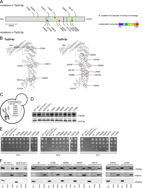
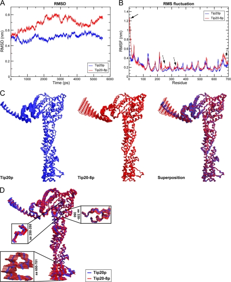
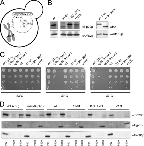
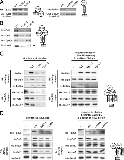
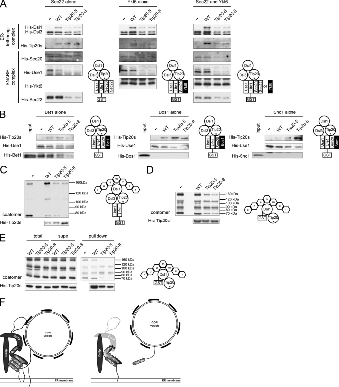
Intracellular transport is largely dependent on vesicles that bud off from one compartment and fuse with the target compartment. The first contact of an incoming vesicle with the target membrane is mediated by tethering factors. The tethering factor responsible for recruiting Golgi-derived vesicles to the ER is the Dsl1 tethering complex, which is comprised of the essential proteins Dsl1p, Dsl3p, and Tip20p. We investigated the role of the Tip20p subunit at the ER by analyzing two mutants, tip20-5 and tip20-8. Both mutants contained multiple mutations that were scattered throughout the TIP20 sequence. Individual mutations could not reproduce the temperature-sensitive phenotype of tip20-5 and tip20-8, indicating that the overall structure of Tip20p might be altered in the mutants. Using molecular dynamics simulations comparing Tip20p and Tip20-8p revealed that some regions, particularly the N-terminal domain and parts of the stalk region, were more flexible in the mutant protein, consistent with its increased susceptibility to proteolysis. Both Tip20-5p and Tip20-8p mutants prevented proper ER trans-SNARE complex assembly in vitro. Moreover, Tip20p mutant proteins disturbed the interaction between Dsl1p and the coatomer coat complex, indicating that the Dsl1p-coatomer interaction could be stabilized or regulated by Tip20p. We provide evidence for a direct role of the Dsl1 complex, in particular Tip20p, in the formation and stabilization of ER SNARE complexes.


Figures

FIGURE 1.
FIGURE 1.
Analysis of Tip20-5 and Tip20-8 mutants. A, sequencing of the tip20-5 and the tip20-8 alleles revealed nine and six amino acid (aa) changes, respectively. An alignment and evaluation of evolutionarily conserved residues for Tip20p was performed using the ConSurf database (41). The conservation scores were normalized and translated to nine color codes, which represent the grade of conservation; 1 is maximum variability and 9 is maximum conservation. The mutations occurring in Tip20-8p (top) and in Tip20-5p (bottom) were mapped onto the linear sequence. Stars indicate mutations that do not occur naturally in sequences of TIP20 homologues in other species. Neither the mutations found in tip20-8 nor the ones identified in tip20-5 cluster on the linear sequence. B, the mutations in Tip20-8p and Tip20-5p are relatively evenly distributed throughout the protein, with some enrichment along the α-helical stalk region of the protein. Mutations occurring in Tip20-8p (left) and in Tip20-5p (right) were incorporated into the x-ray crystal structure of Tip20p (3FHN) using the mutation tool in the Swiss-pdb Viewer (44). The side chain conformations of the mutated residues were regenerated from the backbone structure using the program SCWRL (45). C, schematic drawing of yeast strains expressing variants of Tip20p that contain only one of the mutations identified in tip20-8. D, all single point mutation constructs express Tip20p to a similar extent than the wild-type constructs. Immunoblots of protein extracts from the single point mutation were performed. Detection of Arf1/2p was used as loading control. chr., the chromosomally encoded TIP20 versions. E, none of the single point mutations showed any growth defect at any tested temperature. Growth assays were performed at the indicated temperatures to test the tip20 mutant strains. The tip20-8 strain displays a growth defect at 30 °C and above, whereas the tip20-5 strain only ceases to grow at 37 °C. F, in all strains, most of Tip20p was found in the P13 fraction, which contains mostly ER membranes. A smaller portion of Tip20p was found in the S100 fraction. Subcellular fractionations of the indicated strains were performed and analyzed by immunoblots. Pgk1p was used as a marker for cytosolic proteins, whereas Sec61p served as a marker for ER membranes.
FIGURE 2.
FIGURE 2.
Tip20-8p is more flexible than wild-type Tip20p. A, although Tip20p (blue) behaves rather stably during the molecular dynamics simulation, Tip20-8p (red) as an effect of the mutations shows dramatic changes in the r.m.s.d.. The backbone r.m.s.d. values of each protein structure relative to their starting structures were calculated to estimate the quality and convergence of the molecular dynamics trajectory. B, a striking difference between the Tip20p (blue) and Tip20-8p (red) for the first 25–30 residues (indicated by an arrow) and further differences in the regions of residues 250–260, 330–350, and the C terminus (residues 650–701) (indicated by dashed arrows) could be detected. The sources of the observed differences in r.m.s.d. were determined by computation of the r.m.s.f. Thereby, the movement of each residue in the system with respect to the average position of that residue was calculated for both structures. C, component 2 of the principal component analysis reflects the very large movements in the first 30 N-terminal amino acids. The maximal range as well as intermediate states of the movements for the wild-type Tip20p (left, blue) and the Tip20-8p (middle, red) is shown. On the right, a superposition (Tip20p in blue, Tip20-8p in red) is displayed. D, component 1 of the principle component analysis mirrors the observed fluctuations in the regions of the long α-helical stalk. A superposition of the maximal range as well as intermediate states of the movements for wild-type Tip20p (blue) and Tip20-8p (red) is shown. The boxes represent an enlargement of the regions (amino acids 250–255, 330–350, and 650–701) that displayed the biggest amplitude in movement.
FIGURE 3.
FIGURE 3.
The N terminus of Tip20p is not required for growth or membrane localization. A, schematic drawing of yeast strains expressing variants of Tip20p that contain either a version of Tip20p that is lacking the amino acids 1–81 (Δ1–81) or containing two (I10D,L28E) or one point mutation (V17E), respectively. B, all constructs of N-terminal tip20 variants express Tip20p to a similar extent than the wild-type constructs. Immunoblots of protein extracts from the N-terminal tip20 variants were performed. Detection of Arf1/2p was used as loading control. C, none of the N-terminal tip20 variants showed a growth phenotype. Growth assays were performed at the indicated temperatures using the indicated tip20 variant strains. D, none of the N-terminal tip20 variants showed an aberrant localization of Tip20p. Subcellular fractionations of the indicated strains were performed and analyzed by immunoblots. chr., the chromosomally encoded versions of TIP20.
FIGURE 4.
FIGURE 4.
In vitro assembly of Dsl1 tethering complexes and ER trans-SNARE complexes is affected by Tip20p mutants. A, Tip20p mutants have a lower affinity for Sec20 and Dsl1p than wild-type Tip20p. A GST pulldown was performed with GST-Ds1p or GST-Sec20p and Tip20p variants. B, Dsl1p binding to Tip20-5p or Tip20-8p is drastically decreased. To reconstitute the Dsl1 tethering complex in vitro, pulldown assays were performed as indicated in the graphical representation and analyzed by immunoblotting. Note that Dsl3p and Use1p were added as a complex. Dsl1p and Dsl3p were detected with protein specific antibodies, whereas an anti-His antibody was used to detect Tip20p and Use1p. The arrowhead in the Use1p blot points to the upper band, which is the specific one. C, Tip20p is required for SNARE complex assembly. ER SNARE complexes were reconstituted either in the presence or absence of Dsl1 complex members. Mutant Tip20p proteins strongly reduced the amount of SNARE complexes formed. No effect was observed when the Dsl1 complex members were added after SNARE assembly had occurred. D, Dsl1p does not influence SNARE complex assembly. In vitro pulldown assay were performed as in C, but Dsl1p was omitted. The asterisk under Tip20 in the drawings is meant to illustrate the usage of wild type and mutant versions in the pulldowns.
FIGURE 5.
FIGURE 5.
The assembly of ER SNARE complexes in tip20 mutants is not rescued by alternative v-SNAREs and coatomer binding is affected in the presence of Tip20-5p or Tip20-8p. A, Ykt6p binds to GST-Ufe1p but does not promote SNARE complex assembly. Pulldown assays were performed as indicated in the graphical representation. B, Bet1p, Bos1p, and Snc1p do not improve SNARE complex assembly in the presence of the Tip20p mutants. Pulldown assays were performed as indicated in the graphical representation. C, coatomer binding to mutant Dsl1 complexes is reduced in vitro. To assess whether the coatomer binding function of the Dsl1 complex is affected by the Tip20p mutants, Dsl1 complexes were assembled, and coatomer was added either at the same time or only after Dsl1 complex preassembly. D, Tip20-5 and Tip20-8 negatively regulate the interaction between coatomer and Dsl1p. Tip20p variants and coatomer were incubated with GST-Dsl1p and the binding of the proteins to GST-Dsl1p was determined by immunoblot. E, GST-Dsl1p and Tip20p do not compete for the same binding site on coatomer. Preincubation of Tip20p proteins with coatomer did not change the binding behavior. A pulldown experiment was performed as described in D except that coatomer and the Tip20p proteins were preincubated at 4 °C. To eliminate possible aggregation, the reaction mixture was spun for 15 min at 20,000 × g at 4 °C, and the supernatant was used for the binding reaction to GST-Dsl1p. F, scheme describing the results. For an explanation, see text.

Similar articles

Cited by

References

    1. Bonifacino J. S., Glick B. S. (2004) Cell 116, 153–166 - PubMed
    1. Lee M. C., Miller E. A., Goldberg J., Orci L., Schekman R. (2004) Annu. Rev. Cell Dev. Biol. 20, 87–123 - PubMed
    1. Springer S., Spang A., Schekman R. (1999) Cell 97, 145–148 - PubMed
    1. Barlowe C., Orci L., Yeung T., Hosobuchi M., Hamamoto S., Salama N., Rexach M. F., Ravazzola M., Amherdt M., Schekman R. (1994) Cell 77, 895–907 - PubMed
    1. Letourneur F., Gaynor E. C., Hennecke S., Démollière C., Duden R., Emr S. D., Riezman H., Cosson P. (1994) Cell 79, 1199–1207 - PubMed

Publication types

MeSH terms

Substances

LinkOut - more resources