. 2011 Aug;129(8):1030-41.
doi: 10.1001/archophthalmol.2011.75. Epub 2011 Apr 11.

Development of extraocular muscles requires early signals from periocular neural crest and the developing eye

Affiliations

Development of extraocular muscles requires early signals from periocular neural crest and the developing eye

Brenda L Bohnsack et al. Arch Ophthalmol. 2011 Aug.

Abstract

Objectives: To identify and explain morphologic changes of the extraocular muscles (EOMs) in anophthalmic patients.

Methods: Retrospective medical record review of patients with congenital anophthalmia, using magnetic resonance imaging and intraoperative findings to characterize EOM morphology. We then used molecular biology techniques in zebrafish and chick embryos to determine the relationships among the developing eye, periocular neural crest, and EOMs.

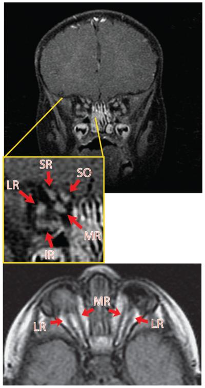
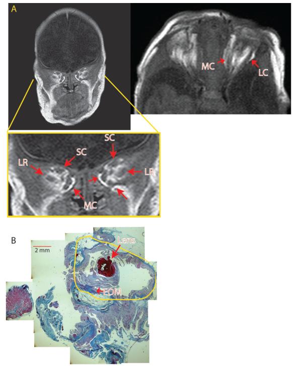
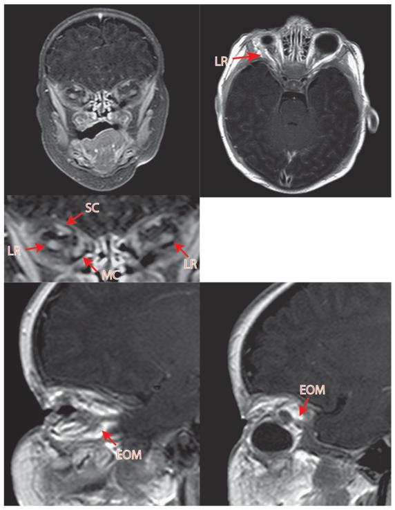
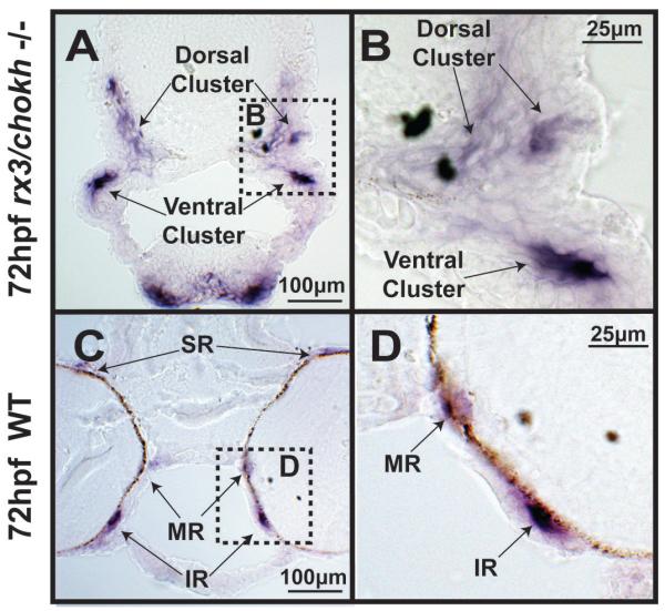
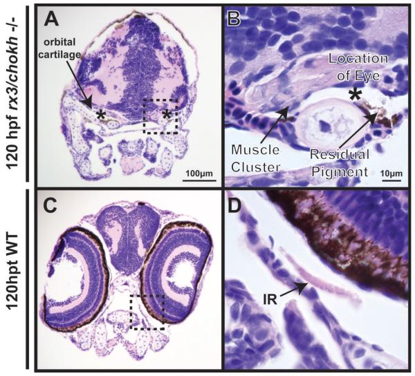
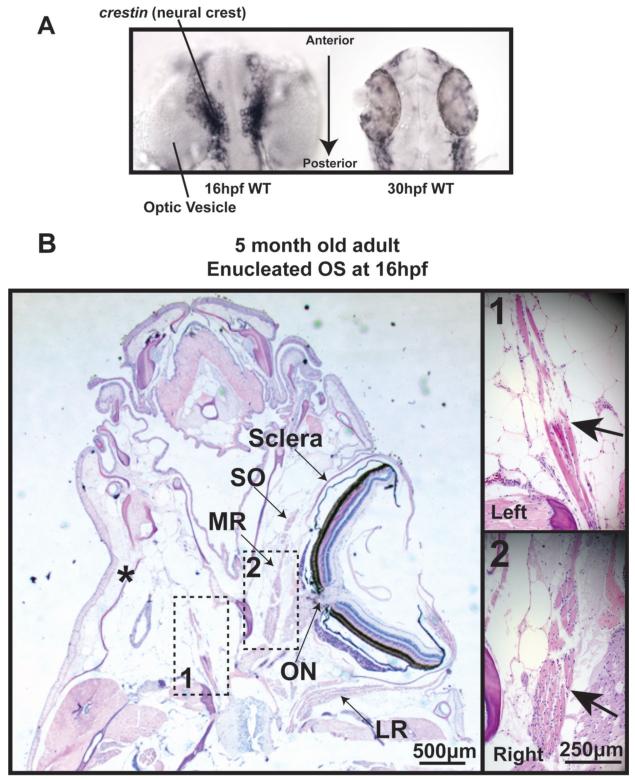
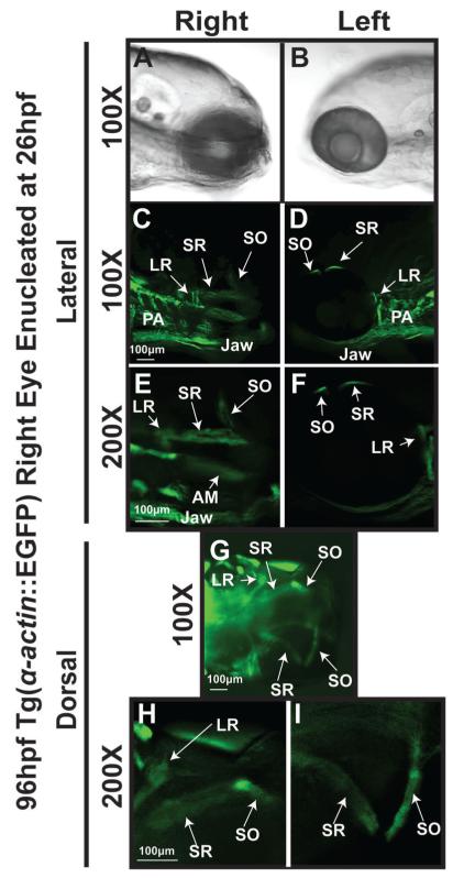
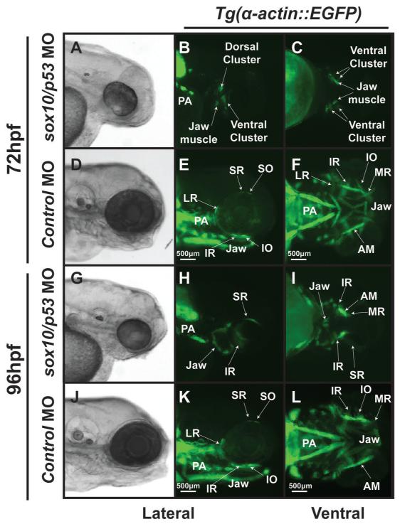
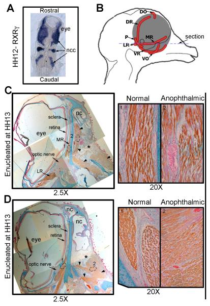
Results: In 3 human patients with bilateral congenital anophthalmia and preoperative orbital imaging, we observed a spectrum of EOM morphologies ranging from indiscernible muscle tissue to well-formed, organized EOMs. Timing of eye loss in zebrafish and chick embryos correlated with the morphology of EOM organization in the orbit (eye socket). In congenitally eyeless Rx3 zebrafish mutants, or following genetic ablation of the cranial neural crest cells, EOMs failed to organize, which was independent of other craniofacial muscle development.

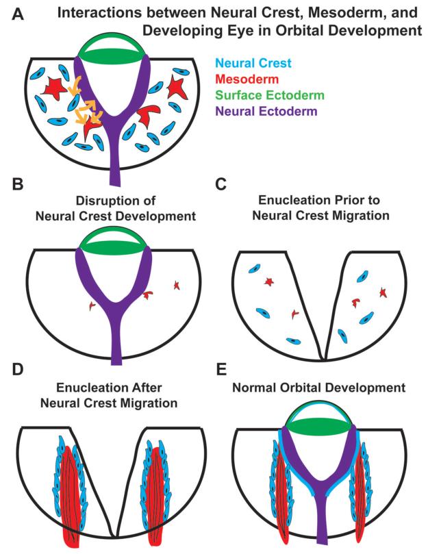
Conclusions: Orbital development is dependent on interactions between the eye, neural crest, and developing EOMs. Timing of the ocular insult in relation to neural crest migration and EOM development is a key determinant of aberrant EOM organization. Additional research will be required to study patients with unilateral and syndromic anophthalmia and assess for possible differences in clinical outcomes of patients with varied EOM morphology.

Clinical relevance: The presence and organization of EOMs in anophthalmic eye sockets may serve as a markers for the timing of genetic or teratogenic insults, improving genetic counseling, and assisting with surgical reconstruction and family counseling efforts.


Figures

Figure 1
Figure 1. Pre-operative MRI of patient 1 with congenital bilateral anophthalmia but well-defined EOMs
MRI of patient 1 reveals the presence of relatively organized EOM groups. Arrows point to the EOMs and EOMs are labeled according to position in the anophthalmic socket. This patient wore small custom socket conformers fitted by an ocularist. Note the absence of an optic nerve. MR, medial rectus; LR, lateral rectus; SR, superior rectus; IR, inferior rectus; SO, superior oblique.
Figure 2
Figure 2. Pre-operative MRI of anophthalmic patient 2 with less well defined XEOMs
A) MRI of patient 2 reveals EOMs that are less defined, with EOM clusters. The coronal image is taken from a more posterior section closer to the muscle origin compared to the MRI in Figure 1. The EOMs of the right orbit are less defined than on the left. B) Section of a cystic eye remnant that was removed from the left socket of patient 2, revealing a small lens (large arrow) within a malformed eye remnant (yellow tracing) surrounded by connective tissue, with irregular EOM insertions onto thickened, poorly formed scleral tissues (small arrow). Intraoperatively, thin EOM remnants were identified. (scale bar = 2 mm.) MC, medial complex; SC, superior complex; LR, lateral rectus.
Figure 3
Figure 3. Pre-operative MRI of patient 3 with congenital bilateral anophthlamia and almost complete lack of EOMs
MRI reveals near total lack of EOMs. A very posterior coronal section is required to identify an EOM cluster, worse on the left, without significant organization. Intraoperatively, EOMs could not be identified on the left side, although fibrovascular connective tissue strands were noted. The hypo-intense round objects in both orbits represent custom external socket conformers fitted by an ocularist.
Figure 4
Figure 4. Genetic ablation of the optic vesicle in Rx3/chokh −/− mutant embryos results in poorly formed EOMs
Wholemount in situ hybridization for myhz2, the gene encoding myosin heavy chain, in Rx3 −/− mutants at 72hpf (A-C) demonstrates the presence of differentiated muscle in the anophthalmic sockets (red arrows). However, the muscles are not properly organized into 6 EOMs, (A-C) as seen in wildtype embryos (G-I).In the Rx3/chokh mutants, muscles of the jaw and pharyngeal arch differentiate and form normally (purple arrows). Schematics of the rx3/chokh mutant (D-F) and wildtype (J-L) show the relation of the EOMs (red) to the eye (blue) and jaw musculature (purple). The dotted line in the lateral view corresponds to the coronal cut shown on the sections shown in Fig 5. SR, superior rectus; MR, medial rectus; LR, lateral rectus; IR, inferior rectus; SO, superior oblique; IO, inferior oblique.
Figure 5
Figure 5. Rx3/chokh −/− mutant embryos express myosin but fail to form distinct EOM structures
Cryosection in situ hybridization for the myosin gene myhz2 of 72hpf Rx3/chokh mutants (A, B) and wildtype (C, D) embryos demonstrates that the EOMs in the Rx3/chokh −/− mutant form dorsal and ventral clusters (double arrows), but do not organize into distinct rectus and oblique muscles. SR, superior rectus; MR, medial rectus; LR, lateral rectus; IR, inferior rectus; SO, superior oblique; IO, inferior oblique.
Figure 6
Figure 6. Rx3/chokh −/− mutant embryos lack orbits with distinct EOMs
H&E staining of coronal sections in Rx3−/− (left) embryos at 120 hpf demonstrate bilateral anophthalmia compared to wildtype controls (right). Rx3 −/− mutants showed cartilage (double arrow) surrounding the shrunken anophthalmic socket (asterik) which contained a small amount of remnant pigment (arrowhead), but no appreciable ocular structures. EOMs (arrows) are present in Rx3 −/− mutants. However, they are thickened and disorganized compared to wildtype control. SR, superior rectus; MR, medial rectus; LR, lateral rectus; IR, inferior rectus; SO, superior oblique; IO, inferior oblique.
Figure 7
Figure 7. Surgical removal of the developing eye alters EOM formation
A) In situ hybridization for crestin expression demonstrates that neural crest is adjacent to the neural tube at 16 hpf (left) and has migrated to peripheral locations by 30 hpf (right). B) H&E staining of a transverse section of 5 month old zebrafish that was enucleated at 16 hpf reveals one rectus muscle close to the origin, but a complete lack of other rectus and oblique muscles in the anophthalmic orbit. (scale bar 250μm)
Figure 8
Figure 8. EOM properly form in orbits that were enucleated following neural crest migration
Tg(α-actin::EGFP) embryo that was enucleated at 26 hpf and imaged at 96hpf (top) demonstrates well formed and distinct EOMs including superior rectus and superior oblique. Contralateral control side (bottom) shows 6 distinct EOMs. SR, superior rectus; SO, superior oblique; LR, lateral rectus; MR, medial rectus.
Figure 9
Figure 9. Inhibition of cranial neural crest development by knockdown of sox10 expression by antisense oligonucleotide morpholinos is required for EOM development
Microinjection of antisense oligonucleotide morpholinos at the 1-2 cell stage (within 30 minutes of fertilization) effectively knocked down the expression of sox10 and inhibited neural crest development.. Knockdown of sox10 in the Tg(α-actin::EGFP) embryos demonstrates poor development of EOMs at 72 (A-C) and 96 hpf (G-I) when compared to control animals (D-F, J-L). MO against p53 was coinjected with the sox10 MO to decrease non-specific (off target) effects, but knockdown of p53 did not alter the EOM phenotype. At 72 hpf, the sox10/p53 morphants showed significant delay in EOM development and overall decreased α-actin expression. By 96 hpf, the sox10/p53 morphants had rudimentary EOM which were severely malformed and not organized around the eye compared to control MO.
Figure 10
Figure 10. Surgical removal of the developing eye in the chick disrupts EOMs
A) Dorsal view in situ hybridization for RXRγ on a HH stage 12 (~E2) chick embryo, showing the close contact between the developing eye and the neural crest cells (blue). B) Schematic representing the chick head of an E14 embryo seen from the midline showing the positioning of the EOMs (red) of the left eye. The blue dotted line suggests the position of the sections in C and D. (C and D) HH stage 40 (E14) chick embryo that was enucleated at HH stage 13 labeled for Myosin (brown), Alcian blue (cartilage- blue) and nuclear fast red (nuclei-red). The position where the eye would have developed is denoted by the *. No retina, optic nerve or sclera has developed in this region. However, extraocular muscles are seen (arrow heads). C) The medial rectus muscle on the anophthalmic side of the embryo (right) has become fully differentiated muscle but is misshapen and has a looser morphology compared to those seen on the control (left) side. D) The other EOMs show reduced organization (right) compared to the control side (left)and individual muscles are no longer discrete and identifiable. Ncc, neural crest cell; MR, medial rectus; VR, ventral rectus; LR, lateral rectus; VO, ventral oblique; P, pyramidalis; DR, dorsal rectus; DO, dorsal oblique; nc, nasal cavity.
Figure 11
Figure 11. Model
The developing eye, EOMs, and migratory cranial neural crest cells form interdependent relationships that are necessary for the proper development of one another (A, E). Disrupting the development of either the eye or the neural crest during early stages of orbital development (B, C) impact one another and also leave a permanent mark on the structural organization of EOMs, whose development requires input signals from both the developing eye and surrounding cranial neural crest cells. Removal of the eye after the migration of the neural crest into the orbit has less effect on EOM development (D).

Comment in

References

    1. Zhou Y, Liu D, Kaminski HJ. Myosin heavy chain expression in mouse extraocular muscle: More complex than expected. Invest Ophthalmol Vis Sci. 2010;51(12):6355–6363. - PMC - PubMed
    1. Kusner LL, Young A, Tjoe S, Leahy P, Kaminski HJ. Perimysial fibroblasts of extraocular muscle, as unique as the muscle fibers. Invest Ophthalmol Vis Sci. 2010;51(1):192–200. - PMC - PubMed
    1. Zeiger U, Khurana TS. Distinctive patterns of microRNA expression in extraocular muscles. Physiol Genomics. 2010;41(3):289–296. - PMC - PubMed
    1. Porter JD, Israel S, Gong B, et al. Distinctive morphological and gene/protein expression signatures during myogenesis in novel cell lines from extraocular and hindlimb muscle. Physiol Genomics. 2006;24(3):264–275. - PubMed
    1. Yu Wai, Man CY, Chinnery PF, Griffiths PG. Extraocular muscles have fundamentally distinct properties that make them selectively vulnerable to certain disorders. Neuromuscul Disord. 2005;1(1):17–23. - PubMed

Publication types

MeSH terms