

Varicella zoster virus ischemic optic neuropathy and subclinical temporal artery involvement
- PMID: 21482932
- PMCID: PMC3238137
- DOI: 10.1001/archneurol.2011.64
Varicella zoster virus ischemic optic neuropathy and subclinical temporal artery involvement
Abstract
Objective: To demonstrate varicella zoster virus (VZV) infection in an asymptomatic extracranial (temporal) artery in a patient with ischemic optic neuropathy produced by VZV vasculopathy in whom the pathological changes were mistakenly identified as giant cell arteritis.
Design: Case report.
Setting: Teaching hospital, pathology and virology laboratory.
Patient: An 80-year-old man with left ophthalmic distribution zoster who developed left ischemic optic neuropathy.
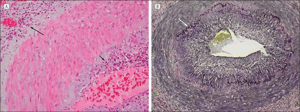
Intervention: An ipsilateral temporal artery biopsy revealed inflammation that was mistakenly identified as giant cell arteritis. The patient was initially treated with steroids but his condition did not improve. When the diagnosis of VZV vasculopathy was confirmed virologically and the patient was treated with intravenous acyclovir, his vision improved.
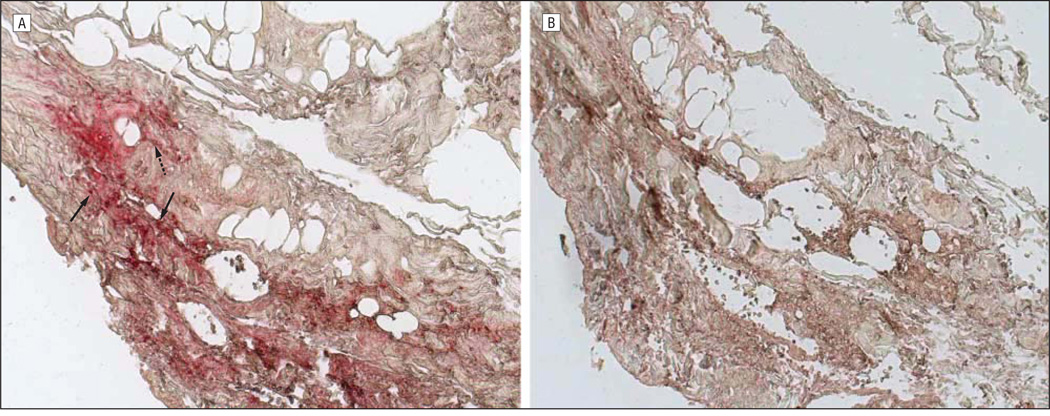
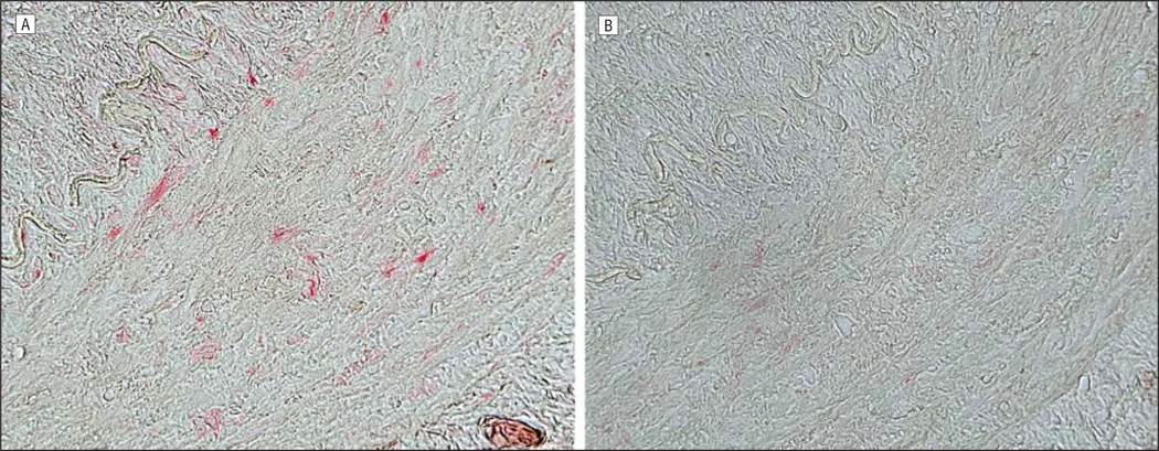
Results: Pathological and virological studies provided proof of VZV vasculopathy in the asymptomatic temporal artery. Varicella zoster virus antigen was abundant in arterial adventitia and scattered throughout the media. With intravenous antiviral therapy, the patient's vision improved.
Conclusion: Although in previously studied patients who died of chronic VZV vasculopathy after 10 to 12 months, VZV antigen was present exclusively in the intima, collective analyses of chronic cases and the asymptomatic VZV-infected temporal artery suggest that virus enters arteries through the adventitia and spreads transmurally to the intima.
Figures
References
-
- Gilden DH, Kleinschmidt-DeMasters BK, Wellish M, Hedley-Whyte ET, Rentier B, Mahalingam R. Varicella zoster virus, a cause of waxing and waning vasculitis: the New England Journal of Medicine case 5-1995 revisited. Neurology. 1996;47:1441–1446. - PubMed
-
- Case Records of the Massachusetts General Hospital. Case records of the Massachusetts General Hospital: weekly clinicopathological exercises case 5-1995: a 73-year-old man with focal brain lesions and peripheral-nerve disease. N Engl J Med. 1995;332(7):452–459. - PubMed
-
- Nagel MA, Forghani B, Mahalingam R, et al. The value of detecting anti-VZV IgG antibody in CSF to diagnose VZV vasculopathy. Neurology. 2007;68(13):1069–1073. - PubMed
-
- Gilden DH, Lipton HL, Wolf JS, et al. Two patients with unusual forms of varicella-zoster virus vasculopathy. N Engl J Med. 2002;347(19):1500–1503. - PubMed
-
- Hall S, Carlin L, Roach ES, McLean WT., Jr Herpes zoster and central retinal artery occlusion. Ann Neurol. 1983;13(2):217–218. - PubMed
Publication types
MeSH terms
Grants and funding
LinkOut - more resources
Full Text Sources
Medical
