

doi: 10.1159/000322101.
Epub 2010 Nov 17.
Orthopox Viruses: Infections in Humans
Affiliations
- PMID: 21483466
- PMCID: PMC3048946
- DOI: 10.1159/000322101
Item in Clipboard
Orthopox Viruses: Infections in Humans
Transfus Med Hemother.
2010.
No abstract available
Figures
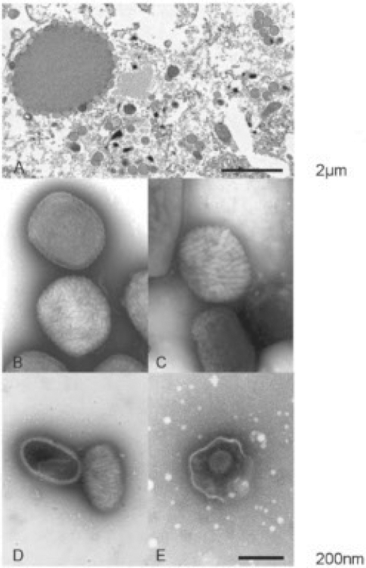
A Electron microscopy: Ultra-thin section of an orthopox virus (OPV)-infected cell. In the cytoplasma (top left) an electron dense 'virus factory' is shown. At the surface of the ‘factory’ virus particles accumulate scattered around the surface. In addition round, immature particles with a clearly visible electron dense envelope can be detected, some with not fully closed envelope. B-E Electron microscopy: Diagnostic negative contrasting of poxvirus and herpes virus. B Two brick-shaped OPV particles with a typical, clearly visible surface structure. The upper particle is penetrated by the contrast stain to show the structure of its lipid membrane while the lower virion only shows its ‘berry’-shaped surface (capsid shape versus mulberry shape of the lower particle). C Molluscum contagiosum virus (MOCV) particles resemble OPV particles in size and shape. However, they also show a slightly different surface structure that can be used for differentiation. D Parapox viruses (PPV; prototype is the orfvirus) are ovoid, show longer, partly parallel surface structures and are markedly smaller than OPV. E Herpes virus from an exsudate used for diagnostic purposes: the capsid of 100 nm diameter with regular shape is still enveloped by the remains of a virus lipid shell. Electron microscopy photography and compilation of the photos by Dr. Andreas Kurth; Robert Koch-Institut, Berlin.
References
-
- Ladnyi ID, Breman JG. Smallpox eradication: progress and problems. Dev Biol Stand. 1978;41:281–290. - PubMed
-
- Geddes AM. The history of smallpox. Clin Dermatol. 2006;24:152–157. - PubMed
-
- WHO Global Commission for the Certification of Smallpox Eradication, editor. The Global Eradication of Smallpox. Final Report of the Global Commission for the Certification of Smallpox Eradication, Geneva, 1979. Geneva: WHO; 1980.
-
- Fenner F, Henderson DA, Arita I, Jezek Z, Ladnyi ID: Smallpox and Its Eradication – World Health Organisation 1988. <ext-link xmlns:xlink="http://www.w3.org/1999/xlink" ext-link-type="uri" xlink:href="http://whqlibdoc.who.int/smallpox/9241561106.pdf">http://whqlibdoc.who.int/smallpox/9241561106.pdf</ext-link>
LinkOut - more resources
Full Text Sources
Other Literature Sources
