. 2011 Mar;7(3):e1001327.
doi: 10.1371/journal.ppat.1001327. Epub 2011 Mar 31.

The lectin receptor kinase LecRK-I.9 is a novel Phytophthora resistance component and a potential host target for a RXLR effector

Affiliations

The lectin receptor kinase LecRK-I.9 is a novel Phytophthora resistance component and a potential host target for a RXLR effector

Klaas Bouwmeester et al. PLoS Pathog. 2011 Mar.

Abstract

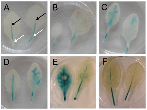
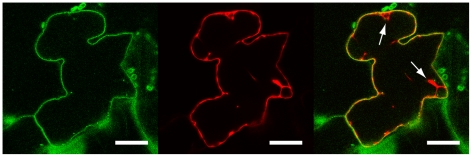
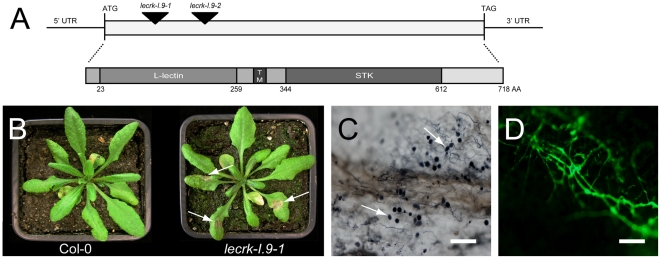
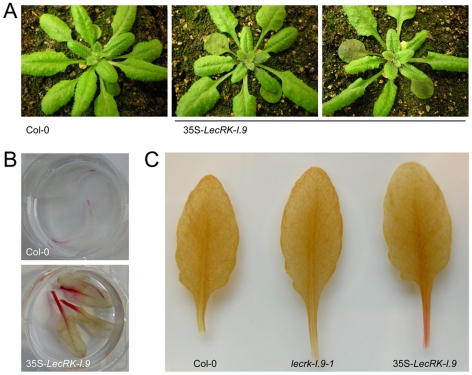
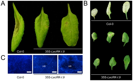
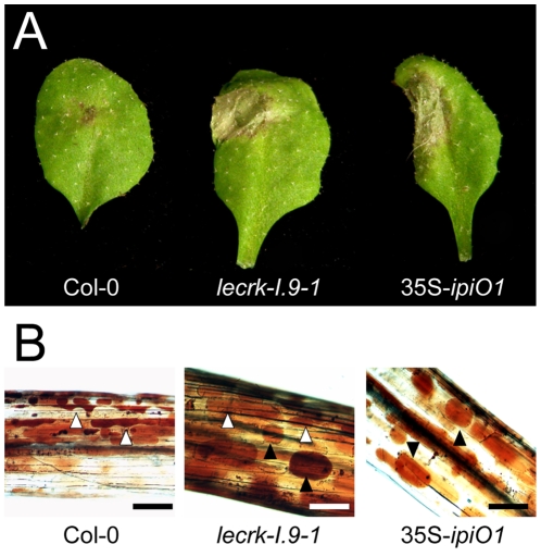
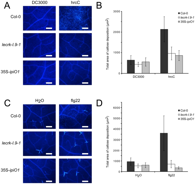
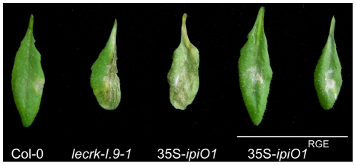
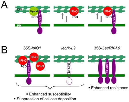
In plants, an active defense against biotrophic pathogens is dependent on a functional continuum between the cell wall (CW) and the plasma membrane (PM). It is thus anticipated that proteins maintaining this continuum also function in defense. The legume-like lectin receptor kinase LecRK-I.9 is a putative mediator of CW-PM adhesions in Arabidopsis and is known to bind in vitro to the Phytophthora infestans RXLR-dEER effector IPI-O via a RGD cell attachment motif present in IPI-O. Here we show that LecRK-I.9 is associated with the plasma membrane, and that two T-DNA insertions lines deficient in LecRK-I.9 (lecrk-I.9) have a 'gain-of-susceptibility' phenotype specifically towards the oomycete Phytophthora brassicae. Accordingly, overexpression of LecRK-I.9 leads to enhanced resistance to P. brassicae. A similar 'gain-of-susceptibility' phenotype was observed in transgenic Arabidopsis lines expressing ipiO (35S-ipiO1). This phenocopy behavior was also observed with respect to other defense-related functions; lecrk-I.9 and 35S-ipiO1 were both disturbed in pathogen- and MAMP-triggered callose deposition. By site-directed mutagenesis, we demonstrated that the RGD cell attachment motif in IPI-O is not only essential for disrupting the CW-PM adhesions, but also for disease suppression. These results suggest that destabilizing the CW-PM continuum is one of the tactics used by Phytophthora to promote infection. As countermeasure the host may want to strengthen CW-PM adhesions and the novel Phytophthora resistance component LecRK-I.9 seems to function in this process.


Conflict of interest statement

The authors have declared that no competing interests exist.

Figures

Figure 1
Figure 1. LecRK-I.9 expression is induced in an incompatible but not in a compatible interaction with P. brassicae.
GUS activity in leaves of a transgenic Arabidopsis line carrying promoter-GUS construct PLecRK-I.9-GUS. (A) In mature non-inoculated leaves GUS activity is localized in the petiole (white arrows) and in veins (black arrows). GUS activity in leaves, 6 days post-inoculation (dpi) with the virulent P. brassicae isolate CBS686.95 (B), the avirulent P. brassicae isolate HH (C), P. infestans IPO-0 (D), 4 dpi with B. cinerea IMI169558 (E), and 2 days after wounding (F).
Figure 2
Figure 2. LecRK-I.9 is localized at the plasma membrane.
Confocal images of a N. benthamiana epidermal cell transiently expressing LecRK-I.9-GFP and mCherry. Single-channel (i.e., GFP left, mCherry middle) and merged fluorescence images (right). Arrows point towards cytoplasmic strands. Scale bars represent 20 µm.
Figure 3
Figure 3. Arabidopsis mutants disrupted in LecRK-I.9 are impaired in resistance to P. brassicae.
(A) Schematic representation of LecRK-I.9 and its gene product. The coding sequence is 2154 nt in length and has one continuous open reading frame. The two mutant lines lecrk-I.9-1 and lecrk-I.9-2 have a T-DNA insertion (black arrowheads) at position 176 and 566, respectively, relative to the translational start. The LecRK-I.9 protein contains an extracellular legume-like lectin domain (L-lectin), a transmembrane motif (TM), and an intracellular serine/threonine protein kinase domain (STK). (B) Col-0 and lecrk-I.9-1 plants 4 days post-inoculation with P. brassicae isolate HH. Arrows point at lesions. Colonization of lecrk-I.9-1 leaves by P. brassicae isolate HH visualized by trypan blue staining (C) and P. brassicae GFP transformant 155 m revealed by UV epifluorescence (D). Arrows in (C) point at sporangia. Scale bar in (C) represents 100 µm and in (D) 200 µm.
Figure 4
Figure 4. LecRK-I.9 overexpression in Arabidopsis leads to changes in morphology and accumulation of anthocyanin and lignin.
In comparison to the recipient line Col-0, 35S-LecRK-I.9 lines have more compact rosettes with smaller and slightly wrinkled leaves (A). Anthocyanin (B) and lignin (C) staining in rosette leaves of Col-0 and 35S-LecRK-I.9 lines.
Figure 5
Figure 5. LecRK-I.9 overexpression in Arabidopsis results in enhanced resistance to P. brassicae.
Arabidopsis leaves inoculated with plugs (A) or zoospores (B and C) of P. brassicae isolate CBS686.95. Pictures were taken 4 days post-inoculation (dpi) (A), 8 dpi (B), and 5 dpi (C). Lesion expansion was observed on Col-0 leaves, but not on leaves from two independent 35S-LecRK-I.9 lines. In (C) leaves were stained for callose (light blue fluorescence). HR; hypersensitive response. Scale bars in (C) represent 50 µm.
Figure 6
Figure 6. Arabidopsis lecrk-I.9 and 35S-ipiO1 lines are phenocopies.
Both lines show (A) gain of susceptibility to P. brassicae, and (B) defects in cell wall integrity. (A) Leaves inoculated with P. brassicae isolate HH 3 days post-inoculation (dpi). (B) Elicitation of plasmolysis in etiolated hypocotyls by treatment with 0.4 M CaCl2. Shown are concave (white arrowheads) and convex (black arrowheads) forms of plasmolysis. In Col-0 only a few cells show convex plasmolysis, in lecrk-I.9 convex shapes occur more frequently whereas in 35S-ipiO1 the majority of cells is convex. Etiolated hypocotyls were stained with neutral red. Scale bars represent 100 µm.
Figure 7
Figure 7. Pathogen- and MAMP-triggered callose deposition is reduced in lecrk-I.9 and 35S-ipiO1 lines.
(A) Arabidopsis leaves stained with aniline blue after infiltration with Pseudomonas syringae DC3000 and hrcC. (B) Average totals of callose deposition (µm2) and associated 95% confidence intervals (CIs) after infiltration with DC3000 and hrcC (n =  >16) (C). Callose deposition after infiltration with flg22. (D) Average totals of callose deposition after treatment with flg22 (µm2) +95% CIs (n =  >10). Scale bars in (A) and (C) represent 50 µm.
Figure 8
Figure 8. The RGD motif in IPI-O is a determinant of the phenotypic changes.
Arabidopsis transgenic lines expressing ipiO1 with a RGD-to-RGE targeted mutation show no gain of susceptibility to P. brassicae HH. Arabidopsis leaves inoculated with P. brassicae strain HH 5 days post-inoculation.
Figure 9
Figure 9. P. infestans effector IPI-O and its putative effector target LecRK-I.9.
(A) Models depicting the membrane-associated LecRK-I.9 and the influence of IPI-O activity on adhesions between cell wall (CW) and plasma membrane (PM). LecRK-I.9 supposedly interacts with RGD-containing extracellular ligands (left panel). Addition of the RGD-containing effector IPI-O alters the CW-PM continuum (middle), and presumably binding of IPI-O to LecRK-I.9 further disrupts this continuum (right). The putative activity of IPI-O inside the plant cell is not included in these models. (B) Observed changes in the Arabidopsis phenocopy lines 35S-ipiO1 and lecrk-I.9 (left and middle panel, respectively), and the LecRK-I.9 overexpressing lines (right panel).

References

    1. Boller T, Felix G. A renaissance of elicitors: Perception of microbe-associated molecular patterns and danger signals by pattern-recognition receptors. Annu Rev Plant Biol. 2009;60:379–407. - PubMed
    1. Hückelhoven R. Cell wall-associated mechanisms of disease resistance and susceptibility. Annu Rev Phytopathol. 2008;45:101–127. - PubMed
    1. Mellersh DG, Heath MC. Plasma membrane-cell wall adhesion is required for expression of plant defense responses during fungal penetration. Plant Cell. 2001;13:413–424. - PMC - PubMed
    1. Ruoslahti E. RGD and other recognition sequences for integrins. Annu Rev Cell Dev Biol. 1996;12:697–715. - PubMed
    1. Watson N, Duncan G, Annan WS, van der Walle CF. A tetravalent RGD ligand for integrin-mediated cell adhesion. J Pharm Pharmacol. 2006;58:959–966. - PubMed

Publication types

MeSH terms

Substances