

Quantification of the optical properties of two-layered turbid media by simultaneously analyzing the spectral and spatial information of steady-state diffuse reflectance spectroscopy
- PMID: 21483612
- PMCID: PMC3072129
- DOI: 10.1364/BOE.2.000914
Quantification of the optical properties of two-layered turbid media by simultaneously analyzing the spectral and spatial information of steady-state diffuse reflectance spectroscopy
Abstract
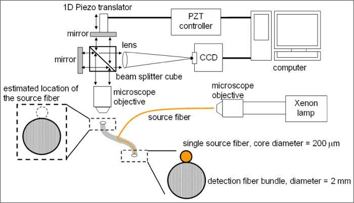
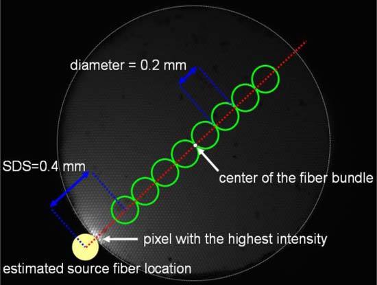
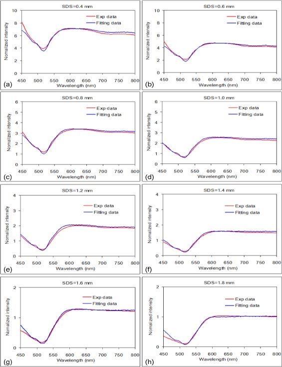
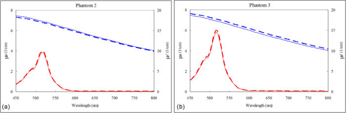
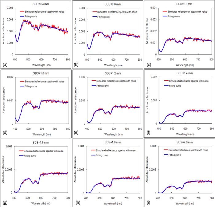
We applied hyperspectral imaging to measure spatially-resolved diffuse reflectance spectra in the visible range and an iterative inversion method based on forward Monte Carlo modeling to quantify optical properties of two-layered tissue models. We validated the inversion method using spectra experimentally measured from liquid tissue mimicking phantoms with known optical properties. Results of fitting simulated data showed that simultaneously considering the spatial and spectral information in the inversion process improves the accuracies of estimating the optical properties and the top layer thickness in comparison to methods fitting reflectance spectra measured with a single source-detector separation or fitting spatially-resolved reflectance at a single wavelength. Further development of the method could improve noninvasive assessment of physiological status and pathological conditions of stratified squamous epithelium and superficial stroma.
Keywords: (110.4234) Multispectral and hyperspectral imaging; (170.3660) Light propagation in tissues; (170.6510) Spectroscopy, tissue diagnostics; (170.7050) Turbid media; (300.6550) Spectroscopy, visible.
Figures
References
-
- McGee S., Mirkovic J., Mardirossian V., Elackattu A., Yu C. C., Kabani S., Gallagher G., Pistey R., Galindo L., Badizadegan K., Wang Z., Dasari R., Feld M. S., Grillone G., “Model-based spectroscopic analysis of the oral cavity: impact of anatomy,” J. Biomed. Opt. 13(6), 064034 (2008). 10.1117/1.2992139 - DOI - PMC - PubMed
-
- Bargo P. R., Prahl S. A., Goodell T. T., Sleven R. A., Koval G., Blair G., Jacques S. L., “In vivo determination of optical properties of normal and tumor tissue with white light reflectance and an empirical light transport model during endoscopy,” J. Biomed. Opt. 10(3), 034018 (2005). 10.1117/1.1921907 - DOI - PubMed
LinkOut - more resources
Full Text Sources