. 2011 Mar 18;2(4):915-26.
doi: 10.1364/BOE.2.000915.

Label-free high-resolution imaging of prostate glands and cavernous nerves using coherent anti-Stokes Raman scattering microscopy

Label-free high-resolution imaging of prostate glands and cavernous nerves using coherent anti-Stokes Raman scattering microscopy

Liang Gao et al. Biomed Opt Express. .

Abstract

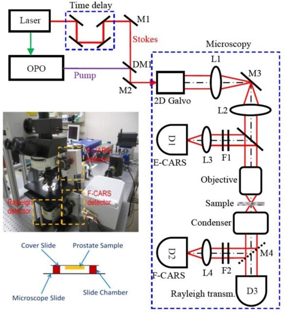
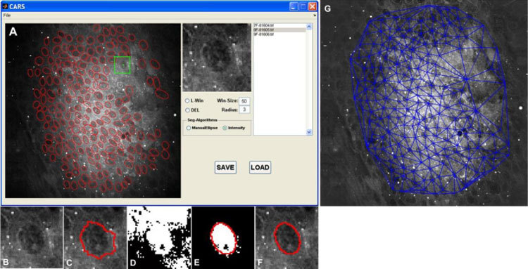
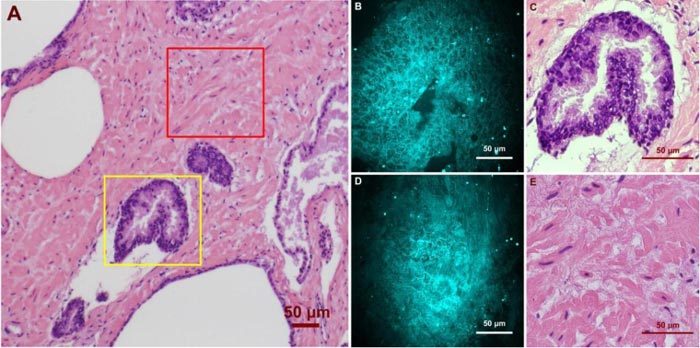
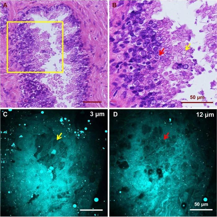
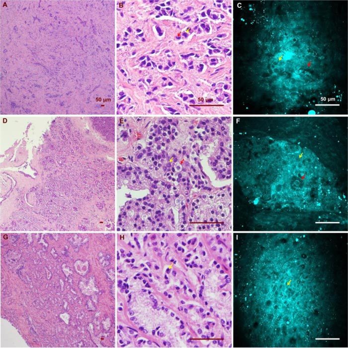
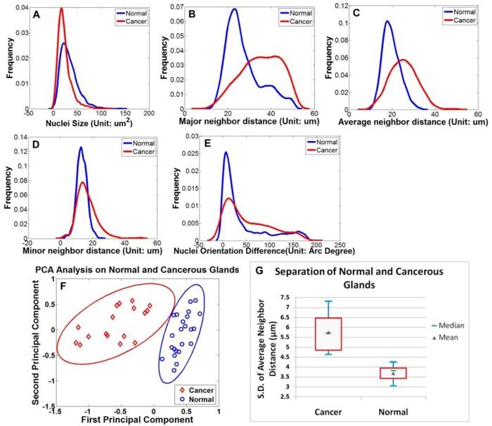
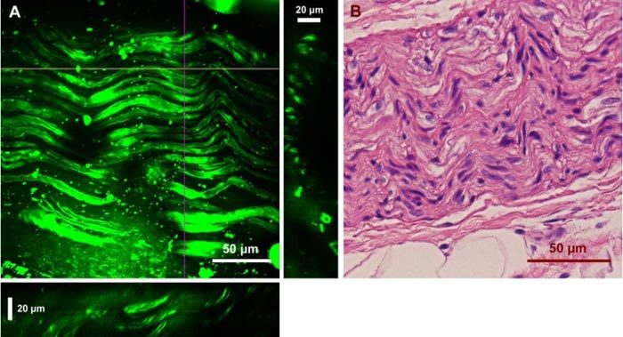
A custom built coherent anti-Stokes Raman scattering (CARS) microscope was used to image prostatic glands and nerve structures from 17 patients undergoing radical prostatectomy. Imaging of glandular and nerve structures showed distinctive cellular features that correlated to histological stains. Segmentation of cell nucleus was performed to establish a cell feature-based model to separate normal glands from cancer glands. In this study, we use a single parameter, average cell neighbor distance based on CARS imaging, to characterize normal and cancerous glandular structures. By combining CARS with our novel classification model, we are able to characterize prostate glandular and nerve structures in a manner that potentially enables real-time, intra-operative assessment of surgical margins and neurovascular bundles. As such, this method could potentially improve outcomes following radical prostatectomy.

Keywords: (170.1610) Clinical applications; (170.3880) Medical and biological imaging; (170.4580) Optical diagnostics for medicine; (180.4315) Nonlinear microscopy.


Figures

Fig. 1
Fig. 1
Schematic of the CARS microscopy system. The pump (816.7 nm) and Stokes (1,064 nm) beams are overlapped, both temporally and spatially, and delivered into a microscopy system. A dichroic mirror is used to separate CARS signals from excitation lasers for detection. D: PMT detector; DM: dichroic mirror; F: filter; L: lens; M: mirror
Fig. 5
Fig. 5
Overview of the nuclear segmentation process and the resulting Delaunay Triangulation graph. (A) Snapshot of graphic user interface of CARS image analysis software; Red ellipses are the delineated cell boundaries. The green star represents one of the user-selected points, while the green square box indicates the image patch which will be processed; intermediate results: (B) targeted nucleus; (C) watershed segmentation algorithm; (D) local thresholding; (E) Ellipse fitting; (F) final result. (G) Delaunay Triangulation on the segmented image.
Fig. 2
Fig. 2
Ex vivo images of human prostate gland and stroma structures using CARS and H&E stain. (A) A low magnification image shows the position of the imaged glandular (Yellow Square) and stroma (Red Square) areas. (B)/(C) high magnification CARS/H&E images of the prostate gland marked in (A). (D)/(E) high magnification CARS/H&E images of the stroma region marked in (A).
Fig. 3
Fig. 3
CARS images of glandular epithelium structures at different depths and H&E images from a similar gland. (A) H&E image of a normal gland showing the appearance of glandular epithelial cells from different imaging planes. (B) Higher magnification image of the highlighted region in (A) showing different appearance of epithelial cells caused by variation in focal planes. Red and yellow arrows point to cells with and without a visible nucleus, respectively. CARS images from a similar gland show epithelial cells as polygons (C) at a shallow plane and with visible nuclei at a deeper plane (D). Cells indicated by the red and yellow arrows in (C) and (D) have structures comparable to those marked in (B).
Fig. 4
Fig. 4
CARS and H&E images of cancerous glands from three patients. (A)(B)(C) Low/ high magnification H&E images and a CARS image from the first patient (Media 1). Red and yellow arrows point to an enlarged epithelial cell and a distinctive cell nucleolus, respectively. (D)(E)(F) Low/ high magnification H&E images and a CARS image from the same patient (Media 2). Red and yellow arrows point to an enlarged epithelial cell containing prominent secretory material in the cytoplasm and a cancerous epithelial cell of relatively normal size. (G)(H)(I) Low/high H&E and CARS results from the third patient, who possesses significant nuclear pleomorphism (Media 3). Yellow arrow points to a distinctive cell nucleolus.
Fig. 6
Fig. 6
Feature extraction and PCA analysis. (A)-(E) Distribution of the five extracted features of the normal (blue) and cancer group (red); (F) Following PCA analysis, the spatial distribution of 23 normal sample sets and 17 cancer sample sets are plotted in the top two principal components space. The eclipses around the data points are 95% confidence intervals for normal (red) and cancer (blue) groups, showing a significant separation between the two groups. (G) Box plot of normal and cancer groups using a single-feature standard deviation of average cell neighbor distance. Blue bars represent samples’ maximum and minimum, while red quadrangles indicate higher and lower quartiles of the samples.
Fig. 7
Fig. 7
Ex vivo images of a human cavernous nerve using CARS and the H&E stained structure of the same nerve. (A) Two-dimensional projection of 45 images from a z-stack with a step size of 1 μm. YZ and XZ cross sections are presented in the right and bottom panels, which were constructed from the depth stack along the purple and yellow lines, respectively. (C) H&E image of the same nerve.

References

    1. W. International Agency for Research on Cancer, “Cancer Incidence and Mortality Worldwide in 2008” (2008), http://globocan.iarc.fr/
    1. Su L. M., “Robot-assisted radical prostatectomy: advances since 2005,” Curr. Opin. Urol. 20(2), 130–135 (2010).10.1097/MOU.0b013e328336257a - DOI - PubMed
    1. Le C. Q., Gettman M. T., “Laparoscopic and robotic radical prostatectomy,” Expert Rev. Anticancer Ther. 6(7), 1003–1011 (2006).10.1586/14737140.6.7.1003 - DOI - PubMed
    1. Gontero P., Kirby R. S., “Nerve-sparing radical retropubic prostatectomy: techniques and clinical considerations,” Prostate Cancer Prostatic Dis. 8(2), 133–139 (2005).10.1038/sj.pcan.4500781 - DOI - PubMed
    1. Grossfeld G. D., Chang J. J., Broering J. M., Miller D. P., Yu J., Flanders S. C., Henning J. M., Stier D. M., Carroll P. R., “Impact of positive surgical margins on prostate cancer recurrence and the use of secondary cancer treatment: data from the CaPSURE database,” J. Urol. 163(4), 1171–1177, quiz 1295 (2000).10.1016/S0022-5347(05)67716-6 - DOI - PubMed

LinkOut - more resources