

Tooth enamel proteins enamelin and amelogenin cooperate to regulate the growth morphology of octacalcium phosphate crystals
- PMID: 21483648
- PMCID: PMC3072691
- DOI: 10.1021/cg100696r
Tooth enamel proteins enamelin and amelogenin cooperate to regulate the growth morphology of octacalcium phosphate crystals
Abstract
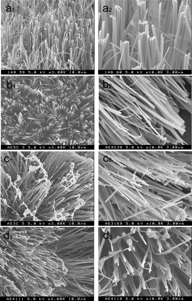
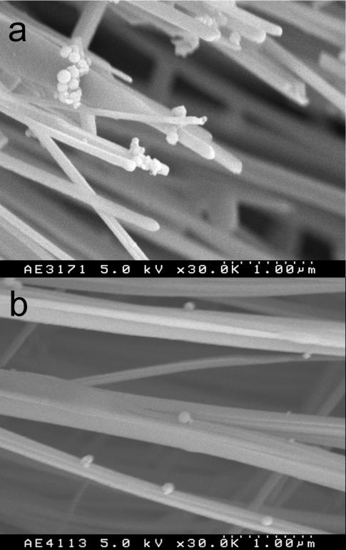
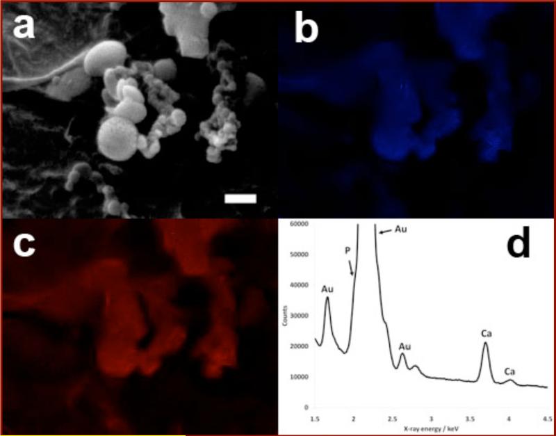
To examine the hypothetical cooperative role of enamelin and amelogenin in controlling the growth morphology of enamel crystals in the post-secretory stage, we applied a cation selective membrane system for the growth of octacalcium phosphate (OCP) in the truncated recombinant porcine amelogenin (rP148) with and without the 32kDa enamelin fragment. Enamelin alone inhibited the growth in the c-axis direction more than rP148, yielding OCP crystals with the smallest aspect ratio of all conditions tested. When enamelin was added to the amelogenin "gel-like matrix", the inhibitory action of the protein mixture on the growth of OCP in the c-axis direction was diminished, while that in the b-axis direction was increased. As a result, the length to width ratio (aspect ratio) of OCP crystal was markedly increased. Addition of enamelin to amelogenin enhanced the potential of amelogenin to stabilize the amorphous calcium phosphate (ACP) transient phase. The ratio of enamelin and amelogenin was crucial for stabilization of ACP and the growth of OCP crystals with larger aspect ratio. The cooperative regulatory action of enamelin and amelogenin was attained, presumably, through co-assembling of enamelin and amelogenin. These results have important implications in understanding the growth mechanism of enamel crystals with large aspect ratio.
Figures


Similar articles
-
Amelogenin-enamelin association in phosphate-buffered saline.Eur J Oral Sci. 2011 Dec;119 Suppl 1(Suppl 1):351-6. doi: 10.1111/j.1600-0722.2011.00916.x. Eur J Oral Sci. 2011. PMID: 22243267 Free PMC article.
-
The cooperation of enamelin and amelogenin in controlling octacalcium phosphate crystal morphology.Cells Tissues Organs. 2011;194(2-4):194-8. doi: 10.1159/000324208. Epub 2011 Apr 28. Cells Tissues Organs. 2011. PMID: 21525716 Free PMC article.
-
Elongated growth of octacalcium phosphate crystals in recombinant amelogenin gels under controlled ionic flow.J Dent Res. 2002 Jan;81(1):69-73. doi: 10.1177/002203450208100115. J Dent Res. 2002. PMID: 11820371
-
Assembly of amelogenin proteolytic products and control of octacalcium phosphate crystal morphology.Connect Tissue Res. 2003;44 Suppl 1:58-64. Connect Tissue Res. 2003. PMID: 12952175 Review.
-
The developing enamel matrix: nature and function.Eur J Oral Sci. 1998 Jan;106 Suppl 1:282-91. doi: 10.1111/j.1600-0722.1998.tb02188.x. Eur J Oral Sci. 1998. PMID: 9541238 Review.
Cited by
-
Matrix metalloproteinase-20 mediates dental enamel biomineralization by preventing protein occlusion inside apatite crystals.Biomaterials. 2016 Jan;75:260-270. doi: 10.1016/j.biomaterials.2015.10.031. Epub 2015 Oct 22. Biomaterials. 2016. PMID: 26513418 Free PMC article.
-
Amelogenin-enamelin association in phosphate-buffered saline.Eur J Oral Sci. 2011 Dec;119 Suppl 1(Suppl 1):351-6. doi: 10.1111/j.1600-0722.2011.00916.x. Eur J Oral Sci. 2011. PMID: 22243267 Free PMC article.
-
The cooperation of enamelin and amelogenin in controlling octacalcium phosphate crystal morphology.Cells Tissues Organs. 2011;194(2-4):194-8. doi: 10.1159/000324208. Epub 2011 Apr 28. Cells Tissues Organs. 2011. PMID: 21525716 Free PMC article.
-
Enamel synthesis explained.Proc Natl Acad Sci U S A. 2020 Sep 8;117(36):21847-21848. doi: 10.1073/pnas.2014394117. Epub 2020 Aug 18. Proc Natl Acad Sci U S A. 2020. PMID: 32817483 Free PMC article. No abstract available.
-
Control of Calcium Phosphate Nucleation and Transformation through Interactions of Enamelin and Amelogenin Exhibits the "Goldilocks Effect".Cryst Growth Des. 2018 Dec 5;18(12):7391-7400. doi: 10.1021/acs.cgd.8b01066. Epub 2018 Oct 22. Cryst Growth Des. 2018. PMID: 32280310 Free PMC article.
References
-
- Lowenstam HA, Weiner S. On Biomineralization. Oxford University Press; New York: 1989.
-
- Moradian-Oldak J, Paine ML. In: Metal Ions In Life Sciences. Astrid Sigel, Sigel H, Sigel RKO., editors. Vol. 4. John Wiley & Sons, Ltd; Chichester: 2008. pp. 507–546. Biomineralization. From Nature to Application.
-
- Veis A. Science. 2005;307:1419–1420. - PubMed
-
- Fearnhead RW. Nature. 1960;189:509. - PubMed
-
- Ronnholm E. J. Ultrastruct. Res. 1962;6:249–303. - PubMed
Grants and funding
LinkOut - more resources
Full Text Sources
Research Materials