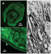
Characterization of schistosome tegumental alkaline phosphatase (SmAP)
- PMID: 21483710
- PMCID: PMC3071363
- DOI: 10.1371/journal.pntd.0001011
Characterization of schistosome tegumental alkaline phosphatase (SmAP)
Abstract
Schistosomes are parasitic platyhelminths that currently infect over 200 million people globally. The parasites can live for years in a putatively hostile environment - the blood of vertebrates. We have hypothesized that the unusual schistosome tegument (outer-covering) plays a role in protecting parasites in the blood; by impeding host immunological signaling pathways we suggest that tegumental molecules help create an immunologically privileged environment for schistosomes. In this work, we clone and characterize a schistosome alkaline phosphatase (SmAP), a predicted ∼60 kDa glycoprotein that has high sequence conservation with members of the alkaline phosphatase protein family. The SmAP gene is most highly expressed in intravascular parasite life stages. Using immunofluorescence and immuno-electron microscopy, we confirm that SmAP is expressed at the host/parasite interface and in internal tissues. The ability of living parasites to cleave exogenous adenosine monophosphate (AMP) and generate adenosine is very largely abolished when SmAP gene expression is suppressed following RNAi treatment targeting the gene. These results lend support to the hypothesis that schistosome surface enzymes such as SmAP could dampen host immune responses against the parasites by generating immunosuppressants such as adenosine to promote their survival. This notion does not rule out other potential functions for the adenosine generated e.g. in parasite nutrition.
Conflict of interest statement
The authors have declared that no competing interests exist.
Figures


Similar articles
-
Schistosome immunomodulators.PLoS Pathog. 2021 Dec 30;17(12):e1010064. doi: 10.1371/journal.ppat.1010064. eCollection 2021 Dec. PLoS Pathog. 2021. PMID: 34969052 Free PMC article. Review.
-
Intravascular Schistosoma mansoni Cleave the Host Immune and Hemostatic Signaling Molecule Sphingosine-1-Phosphate via Tegumental Alkaline Phosphatase.Front Immunol. 2018 Jul 30;9:1746. doi: 10.3389/fimmu.2018.01746. eCollection 2018. Front Immunol. 2018. PMID: 30105025 Free PMC article.
-
Vitamin B6 Acquisition and Metabolism in Schistosoma mansoni.Front Immunol. 2021 Feb 4;11:622162. doi: 10.3389/fimmu.2020.622162. eCollection 2020. Front Immunol. 2021. PMID: 33613557 Free PMC article.
-
Schistosomes can hydrolyze proinflammatory and prothrombotic polyphosphate (polyP) via tegumental alkaline phosphatase, SmAP.Mol Biochem Parasitol. 2019 Sep;232:111190. doi: 10.1016/j.molbiopara.2019.111190. Epub 2019 May 30. Mol Biochem Parasitol. 2019. PMID: 31154018 Free PMC article.
-
Schistosoma mansoni and the purinergic halo.Trends Parasitol. 2022 Dec;38(12):1080-1088. doi: 10.1016/j.pt.2022.09.001. Epub 2022 Sep 28. Trends Parasitol. 2022. PMID: 36182536 Free PMC article. Review.
Cited by
-
Immunization with tegument nucleotidases associated with a subcurative praziquantel treatment reduces worm burden following Schistosoma mansoni challenge.PeerJ. 2013 Apr 2;1:e58. doi: 10.7717/peerj.58. Print 2013. PeerJ. 2013. PMID: 23638396 Free PMC article.
-
Can antibody conjugated nanomicelles alter the prospect of antibody targeted therapy against schistosomiasis mansoni?PLoS Negl Trop Dis. 2023 Dec 1;17(12):e0011776. doi: 10.1371/journal.pntd.0011776. eCollection 2023 Dec. PLoS Negl Trop Dis. 2023. PMID: 38039267 Free PMC article.
-
Pyrimidine metabolism in schistosomes: A comparison with other parasites and the search for potential chemotherapeutic targets.Comp Biochem Physiol B Biochem Mol Biol. 2017 Nov;213:55-80. doi: 10.1016/j.cbpb.2017.07.001. Epub 2017 Jul 21. Comp Biochem Physiol B Biochem Mol Biol. 2017. PMID: 28735972 Free PMC article. Review.
-
Generation of Immune Modulating Small Metabolites-Metabokines-By Adult Schistosomes.Pathogens. 2025 May 24;14(6):526. doi: 10.3390/pathogens14060526. Pathogens. 2025. PMID: 40559534 Free PMC article.
-
Schistosome immunomodulators.PLoS Pathog. 2021 Dec 30;17(12):e1010064. doi: 10.1371/journal.ppat.1010064. eCollection 2021 Dec. PLoS Pathog. 2021. PMID: 34969052 Free PMC article. Review.
References
-
- King CH, Dangerfield-Cha M. The unacknowledged impact of chronic schistosomiasis. Chronic Illn. 2008;4:65–79. - PubMed
-
- King CH, Dickman K, Tisch DJ. Reassessment of the cost of chronic helmintic infection: a meta-analysis of disability-related outcomes in endemic schistosomiasis. Lancet. 2005;365:1561–1569. - PubMed
-
- Keating J, Wilson R, Skelly P. No overt cellular inflammation around intravascular schistosomes in vivo. J Parasitol. 2006;92:1365–1369. - PubMed
-
- Morris GP, Threadgold LT. Ultrastructure of the tegument of adult Schistosoma mansoni. J Parasitol. 1968;54:15–27. - PubMed
-
- Smith JH, Reynolds ES, Von Lichtenberg F. The integument of Schistosoma mansoni. Am J Trop Med Hyg. 1969;18:28–49. - PubMed
Publication types
MeSH terms
Substances
Associated data
- Actions
Grants and funding
LinkOut - more resources
Full Text Sources

