. 2011 Mar 29:5:21.
doi: 10.3389/fnana.2011.00021. eCollection 2011.

The evolution of dopamine systems in chordates

Affiliations

The evolution of dopamine systems in chordates

Kei Yamamoto et al. Front Neuroanat. .

Abstract

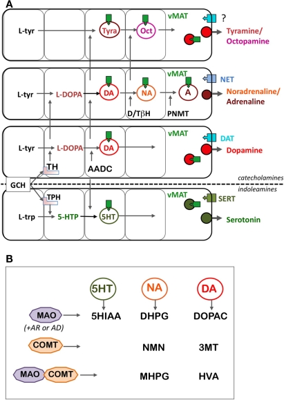
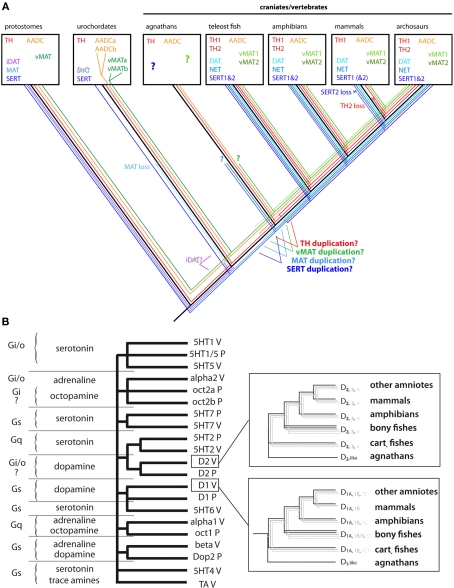
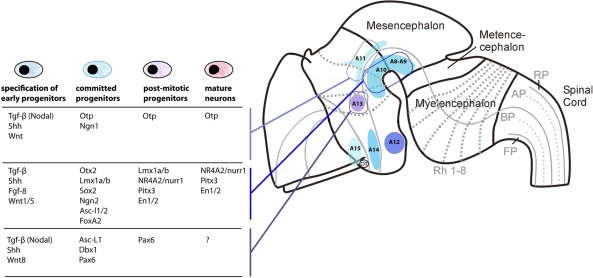
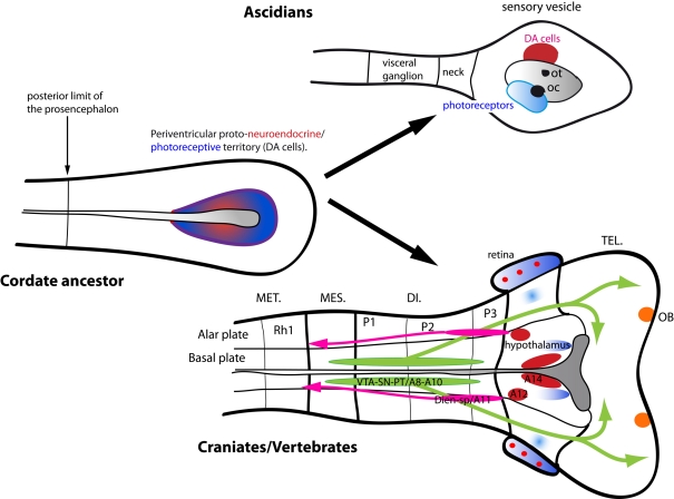
Dopamine (DA) neurotransmission in the central nervous system (CNS) is found throughout chordates, and its emergence predates the divergence of chordates. Many of the molecular components of DA systems, such as biosynthetic enzymes, transporters, and receptors, are shared with those of other monoamine systems, suggesting the common origin of these systems. In the mammalian CNS, the DA neurotransmitter systems are diversified and serve for visual and olfactory perception, sensory-motor programming, motivation, memory, emotion, and endocrine regulations. Some of the functions are conserved among different vertebrate groups, while others are not, and this is reflected in the anatomical aspects of DA systems in the forebrain and midbrain. Recent findings concerning a second tyrosine hydroxylase gene (TH2) revealed new populations of DA-synthesizing cells, as evidenced in the periventricular hypothalamic zones of teleost fish. It is likely that the ancestor of vertebrates possessed TH2 DA-synthesizing cells, and the TH2 gene has been lost secondarily in placental mammals. All the vertebrates possess DA cells in the olfactory bulb, retina, and in the diencephalon. Midbrain DA cells are abundant in amniotes while absent in some groups, e.g., teleosts. Studies of protochordate DA cells suggest that the diencephalic DA cells were present before the divergence of the chordate lineage. In contrast, the midbrain cell populations have probably emerged in the vertebrate lineage following the development of the midbrain-hindbrain boundary. The functional flexibility of the DA systems, and the evolvability provided by duplication of the corresponding genes permitted a large diversification of these systems. These features were instrumental in the adaptation of brain functions to the very variable way of life of vertebrates.

Keywords: forebrain; gene duplication; hypothalamus; monoamine receptors; monoamine transporters; protochordates; tyrosine hydroxylase; vertebrates.


Figures

Figure 1
Figure 1
Schematic representation of the metabolic (A) and catabolic (B) pathways of DA and other monoamines. (A) The biosynthesis of DA is highly modular and shares several molecular components with biosynthetic pathways of other monoamines. Catecholamines are synthesized from the aromatic amino acid l-tyrosine, and indoleamines synthesized from the aromatic amino acid l-tryptophan, but TH and TPH display many common characteristics, including the same co-factor (tetrahydrobiopterin, pinkish square under TH and TPH). Tyramine and octopamine come from the same pathways than the other catecholamines, without the primary action of TH. Then, AADC and vMAT are components shared by all the pathways, including indoleamines. Finally, the membrane transporters, DAT, NET, and SERT, responsible for the re-uptake of the monoamines are more specific of the different monoamine pathways based on their cell-expression pattern, although their functional specificity is weak, especially for the DAT and NET (see text). (B) The catabolic enzymes are also essentially shared by all the monoamines. MAO is an intracellular enzyme, whose direct metabolites are rapidly transformed by aldehyde reductases (AR) and aldehyde dehydrogenase (AD) in compounds (5HIAA for serotonin, DHPG for noradrenaline, and DOPAC for dopamine) easy to assay and which reflect the utilization of the transmitters. The effect of COMT, combined to that of MAO, provides metabolites assayable in body fluid such as CSF, blood, or urine, reflecting preferentially the utilization of monoamines at the periphery of the body. Abbreviations: 3MT, 3-methoxytyramine; 5HIAA, 5-hydroxy-acetic acid; 5-HTP, 5-hydroxytryptophan; AADC, aromatic amino acid decarboxylase; AD, aldehyde dehydrogenase; AR, aldehyde reductase; COMT, catechol-O-methyl transferase; DAT, dopamine transporter; DHPG, 3,4-dihydroxyphenyl-ethylene-glycol; DOPAC, 3,4-dihydroxyphenylacetate; D/TβH, dopamine/tyramine β-hydroxylase; GCH, GTP cyclohydrolase; HVA, homovanillic acid; l-trp, l-tryptophan; l-tyr, l-tyrosine; MAO, monoamine oxidase; MHPG, 3-methoxy-4-hydroxyphenyl-ethylene-glycol; NET, noradrenaline transporter; NMN, normetanephrine; Oct, octopamine; PNMT, phenylethanolamine-N-methyl transferase; SERT, serotonine transporter; TH, tyrosine hydroxylase; Tyra, tyramine; vMAT, vesicular monoamine transporter.
Figure 2
Figure 2
Evolution of the molecular components of the monoaminergic systems in chordates. (A) Protochordates have all the basic molecular components of the monoamine pathways found in vertebrates. In urochordates, both MAT and iDAT do not exist but since MAT is present in amphioxus, it may have been lost specifically in urochordates. The loss of iDAT may have occurred earlier since it is not present in amphioxus. In vertebrates, MAT has been duplicated to provide DAT and NET, which are not thus orthologous to invertebrate iDAT but to invertebrate MAT. Similarly, SERT has been duplicated in jawed vertebrates. vMAT and AADC have been specifically duplicated in the urochordate lineage. Close to the emergence of vertebrates, TH, AADC, and vMAT have been duplicated. But since no genomic data are available yet in agnathans, it is currently impossible to known if this duplication took place before or after the emergence of jawed vertebrates. TH2 and SERT2 have been lost in placental mammals. Abbreviations and colors: AADC, aromatic amino acid decarboxylase (orange); iDAT and DAT, invertebrate form (i), and vertebrate form of dopamine transporter (light blue); MAT, monoamine transporter (blue); NET, noradrenaline transporter (blue); SERT, serotonine transporter (marine blue); TH, tyrosine hydroxylase (red); vMAT, vesicular monoamine transporter (green). (B) The molecular phylogeny of monoamine receptor in bilaterian animals reveals that most classes of monoamine receptors predated the origin of chordates and vertebrates. Classes of orthologous receptors in vertebrates and protostomes (most sequences come from ecdysozoan insects and nematodes) are transducing signals in cells via the same G protein (question marks correspond to the cases when the nature of G protein in not known), highlighting one of the major constraint on the conservation of the receptor sequences throughout bilaterian evolution. For example, α1 adrenergic receptors are orthologous to octopamine 1 receptors (Oct1) and α2 adrenergic receptors are orthologous to octopamine 2 receptors (Oct2), but both D1-like and D2-like receptors are also dopaminergic in protostomes. The topology of the tree also shows that receptor classes that bind the same natural ligand (e.g., DA) are not grouped together, suggesting that each class of receptor acquired independently and convergently the ability to bind a given neurotransmitter. Inside the rectangle, a simplified version of the phylogenetical relationships of the D1 and D2 receptor is presented. Three subtypes of receptor exist in each class, with the notable exception of mammals.
Figure 3
Figure 3
Localization of comparable DA cells among in different groups of vertebrates. A sagittal view of the mammalian (mouse), avian (chicken), amphibian (frog), and teleostean (zebrafish) brains showing comparable DAergic nuclei and their projection patterns. Mammalian A9–A14 DA nuclei are shown in different colors (top), and corresponding colors in other vertebrate groups represent comparative (not necessarily being confirmed to be homologous) DA cell populations. TH2 (green) and pretectal (brown) DA cell groups are commonly found in vertebrates except mammals. Midbrain is shaded with dark gray, diencephalon in gray, and telencephalon in light gray in the brain of each species. The approximate positions of prosomeres p1–p3 are indicated as segmentation within the diencephalon. Abbreviations: Cb, cerebellum; Hy, hypothalamus; M, midbrain; NCL, nidopallium caudolaterale; OB, olfactory bulb; PFC, prefrontal cortex; Str, striatum.
Figure 4
Figure 4
Distinct differentiation pathways for different DA cell groups in mouse. The localization of the main DA-synthesizing nuclei is shown on a neuromeric representation of the mouse embryonic brain. Signaling molecules and genes known to be involved in the differentiation of the A11 (first row) A8–A10 DA nuclei (in the midbrain and first basal diencephalic prosomeres; second row) and A13 DA nuclei (in the ventral thalamus; third row) are indicated as a table. Note that DA cells in the three different areas depend on different signaling and transcription factors expression at each step of the cell differentiation, although little is still known in the case of A11 and A13. This observation supports the hypothesis that different DA neuronal systems have been recruited independently, by different transcriptional mechanisms, probably early in vertebrate evolution.
Figure 5
Figure 5
Schematic representation of the hypothetic evolution of DA systems at the protochordate–vertebrate transition. The anterior neural tube of a chordate ancestor contained periventricular photoreceptor cells intermingled with neuroendocrine cells synthesizing dopamine and peptides such as vasopressin or somatostatin. These cells could also be connected to other part of the CNS, in particular a central pattern generator. These cell types are lining the anterior neural ventricle and contact the CSF. A reminiscent but derived situation is found in modern protochordates such as ascidia (superior right part of the schematics) where photoreceptor cells line the ventricle and are adjacent to the DA cells of the sensory vesicle, and which are able to modulate the motor response to light. In craniates/vertebrates, the telencephalon has tremendously increased in size, and the optic vesicle becomes separated from the anterior hypothalamus by bulging out of the neural tube. Several “new” DA systems have been co-opted simultaneously, mostly in the mesencephalon and the basal plate three first prosomeres. These DA cells homologous to those of the SN/VTA project on the dorsal and ventral pallium, representing a major innovation of vertebrates, with strong adaptive properties. The retina comprised several cell types inherited from the protochordate ancestor, including at least photoreceptor cells, pigmented epithelium, and amacrine DA cells.

References

    1. Acampora D., Postiglione M. P., Avantaggiato V., Di Bonito M., Vaccarino F. M., Michaud J., Simeone A. (1999). Progressive impairment of developing neuroendocrine cell lineages in the hypothalamus of mice lacking the Orthopedia gene. Genes Dev. 13, 2787–2800 10.1101/gad.13.21.2787 - DOI - PMC - PubMed
    1. Agathocleous M., Harris W. A. (2009). From progenitors to differentiated cells in the vertebrate retina. Annu. Rev. Cell Dev. Biol. 25, 45–69 - PubMed
    1. Aguayo L. G., Grossie J. (1994). Dopamine inhibits a sustained calcium current through activation of alpha adrenergic receptors and a GTP-binding protein in adult rat sympathetic neurons. J. Pharmacol. Exp. Ther. 269, 503–508 - PubMed
    1. Allen Z. J. N., Waclaw R. R., Colbert M. C., Campbell K. (2007). Molecular identity of olfactory bulb interneurons: transcriptional codes of periglomerular neuron subtypes. J. Mol. Histol. 38, 517–525 10.1007/s10735-007-9115-4 - DOI - PubMed
    1. Andersson E., Jensen J. B., Parmar M., Guillemot F., Bjorklund A. (2006). Development of the mesencephalic dopaminergic neuron system is compromised in the absence of neurogenin 2. Development 133, 507–516 10.1242/dev.02224 - DOI - PubMed

LinkOut - more resources