. 2011 Mar;7(3):e1001351.
doi: 10.1371/journal.pgen.1001351. Epub 2011 Mar 31.

Cancer-associated splicing variant of tumor suppressor AIMP2/p38: pathological implication in tumorigenesis

Affiliations

Cancer-associated splicing variant of tumor suppressor AIMP2/p38: pathological implication in tumorigenesis

Jin Woo Choi et al. PLoS Genet. 2011 Mar.

Abstract

Although ARS-interacting multifunctional protein 2 (AIMP2, also named as MSC p38) was first found as a component for a macromolecular tRNA synthetase complex, it was recently discovered to dissociate from the complex and work as a potent tumor suppressor. Upon DNA damage, AIMP2 promotes apoptosis through the protective interaction with p53. However, it was not demonstrated whether AIMP2 was indeed pathologically linked to human cancer. In this work, we found that a splicing variant of AIMP2 lacking exon 2 (AIMP2-DX2) is highly expressed by alternative splicing in human lung cancer cells and patient's tissues. AIMP2-DX2 compromised pro-apoptotic activity of normal AIMP2 through the competitive binding to p53. The cells with higher level of AIMP2-DX2 showed higher propensity to form anchorage-independent colonies and increased resistance to cell death. Mice constitutively expressing this variant showed increased susceptibility to carcinogen-induced lung tumorigenesis. The expression ratio of AIMP2-DX2 to normal AIMP2 was increased according to lung cancer stage and showed a positive correlation with the survival of patients. Thus, this work identified an oncogenic splicing variant of a tumor suppressor, AIMP2/p38, and suggests its potential for anti-cancer target.


Conflict of interest statement

The authors have declared that no competing interests exist.

Figures

Figure 1
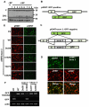
Figure 1. Generation of AIMP2 splicing variant lacking exon 2.
(A) The AIMP2/p38 structural gene consists of four exons encoding a total of 320 aa (NM_006303) with three introns. The 312 aa version of AIMP2 is also reported as U24169. Both forms appear to work similarly for most of our experiments although we do not know the reason for the existence of the two forms at this point. The sequences of exon 1, 2 and 3, and the primers, JTV13, JTV5, JTV11 and DX2-S2 are shown in Figure S1. (B) Expression of AIMP2-F and –DX2 was examined in different lung cells (Normal: WI-26, NL-20; tumorigenic: A549, H322, H460 and H157) by RT-PCR using their specific primers shown above. The pair of the primers, JTV13 and JTV11, generates the two PCR products, one from the transcript of AIMP2-full length (upper band) and the other from the exon 2-lacking transcript (lower band). RT-PCR with the primers, DX2-S2, (specific to the junction site of exon 1 and 3, Figure S1) and JTV5, generates the amplicon only from AIMP2-DX2 transcript. Actin was used as the control. (C) Expression of AIMP2-F and AIMP2-DX2 was also determined in different types of human lung cancer tissues and their normal counterparts by RT-PCR. The primer pairs of JTV-13/JTV-5, and DX2-S2/JTV-5 were used for RT-PCR of AIMP2-F and –DX2, respectively. (D) The expression of AIMP2-F and –DX2 were determined in adenocarcinoma (n = 14) and normal (n = 11) lung tissues by quantitative real-time RT-PCR. The cancer regions were obtained by laser microdissection system from archival formalin-fixed paraffin-embedded (FFPE) patient tissues for RT-PCR as described in experimental procedures. PAPOLA was chosen as the endogenous reference gene for quantitative RT-PCR. The expression results were analyzed by Mann-Whitney test and statistical analyses were achieved using SPSS software (SPSS, Chicago, IL). Each dot represents the expression values of AIMP2-F and -DX2 calibrated to the expression level of PAPOLA. The mean values are shown as lines. **, P<0.01 (E) The larger and smaller amplicons that were generated by RT-PCR with the primers JTV13 and JTV11 were cloned and expressed in AIMP2-deficient MEFs and their expression was determined by Western blotting with monoclonal anti-AIMP2 antibody (#324) that recognizes both forms of AIMP2. Small interference RNAs targeting AIMP2-F and –DX2 were designed and introduced into A549 cells, and their specific effect on the expression of the corresponding transcripts was determined by Western blotting with anti-AIMP2 antibody (right panel).
Figure 2
Figure 2. Splicing variant of AIMP2 is increased by carcinogen-induced mutation.
(A) Normal lung WI-26 cells were incubated in the presence of BPDE for 4 weeks. The surviving colonies were isolated and further cultivated for additional 2 weeks after removal of BPDE to establish stable cell lines. The expression of AIMP2-F and –DX2 in each isolated cell line was determined by Western blotting with anti-AIMP2 antibody. (B) Schematic representation of the EGFP reporter cassette that monitors alternative splicing in cells (pGINT) . Normal splicing process would delete the intervening DNA sequence that is inserted into the open reading frame of EGFP to restore the expression of EGFP. (C) The intervening sequence is inserted into the 146 base position of 717 bp open reading frame of EGFP. The DNA region spanning the 3′ region of intron 1 (630 bp)-exon 2 (207 bp)-intron 2 (426 bp) of AIMP2 was inserted into the open reading frame of EGFP (pGINT-exon 2). If splicing skips exon 2, EGFP will be expressed (upper). However, the normal splicing including exon 2 will break the open reading frame of EGFP (lower). (D) pGINT (left) or pGINT-exon 2 (right) was introduced into HEK293 cells with pRINT (Red fluorescence protein reporter cassette that generates RFP through constitutive splicing intron), and expression of GFP and RFP was observed. (E) pGINT plasmids containing AIMP2 gene with the indicated mutations were tranfected into HEK293 cells and the expression of EGFP was determined. While pGINT empty vector generated EGFP, pGINT containing the wild type exon 2 and flanking intron regions reduced the expression of EGFP. The effect of the indicated mutations on the exon 2 splicing was monitored by the production of EGFP. All the transfectants showed similar degree of RFP, indicating that the difference in EGFP did not result from the variation of transfection efficiency. (F) Expression of the two transcripts (GFP transcripts with and without AIMP2 exon 2 insert) was also determined by RT-PCR.
Figure 3
Figure 3. AIMP2-DX2 compromises pro-apoptotic activity of AIMP2-F.
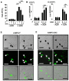
(A) AIMP2-F and –DX2 were transfected into A549 cells and their effects on adriamycin-induced cell death were compared by sub-G1 population using flow cytometry. *: P<0.05. **: P<0.01. A549 cells were transfected with luciferase reporter gene under the control of GADD45 promoter with the indicated amounts of AIMP2-F (B) or AIMP2-DX2 (C), and incubated for additional 12 hours. Adriamycin was treated to the transfectants for 12 hours and luciferase activity was measured by luminometer. (D) MEF cells were transfected with pcDNA3.1 vector and selected by G418 for 2 weeks to establish immortalized cell line. The cells were then transfected with pEGFP-C2 plasmid expressing AIMP2-F. After 24 h, VP-16 (10 µM, Sigma) was added on the dishes and the cells were cultivated for 8 h and the cell morphology was monitored by bright field and fluorescence (489 nm excitation/509 nm emission) microscopy. The images were converged by Metamorph 7.0 imaging program. Bar, 20 µm. (E) EGFP-fused AIMP2-DX2 was also expressed in the same cells and cell morphology was examined in 16 h after VP-16 treatment. Bar, 20 µm.
Figure 4
Figure 4. The effect of AIMP2-DX2 on the interaction of AIMP2-F with p53.
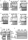
(A) p53 was immunoprecipitated from A549 cells transfected with Myc-AIMP2-F and –DX2, and co-precipitation of the two AIMP2 proteins with p53 was determined with anti-Myc antibody. (B) AIMP2-F and –DX2 were radioactively synthesized by in vitro translation and each protein (20 µl of the in vitro translation reaction) was mixed with GST or GST-p53. GST proteins were precipitated with glutathione-Sepharose beads and co-precipitation of AIMP2-F and –DX2 was determined by autoradiography. (C) Radioactively synthesized AIMP2-F and –DX2 as above were used to see whether the two proteins would compete for the interaction with p53. The indicated amount of AIMP2-DX2 (indicated by microliter volume) was mixed with the fixed amount of AIMP2-F and GST-p53 to see the effect of AIMP2-DX2 on the AIMP2-F binding to p53. (D) Conversely, the indicated amount of AIMP2-F was added to the fixed amount of AIMP2-DX2. (E) MDM2, AIMP2-F and –DX2 were radioactively synthesized as above and the indicated volumes (µl) of AIMP2-F was added to the binding mixture of MDM2 and GST-p53. The mixture was precipitated with glutathione-Sepharose and co-precipitation of MDM2 and AIMP2-F was determined by autoradiography. (F) The same experiment as above was conducted except that AIMP2-DX2 was added at the indicated amounts to the mixture. (G) WI-26 and A549 cells were subjected to UV irradiation and lysed. The extracted proteins were immunoprecipitated with anti-p53 antibody (FL-393), separated by SDS-PAGE and immunoblotted by immunoblotting with the anti-AIMP2 antibody. (H) A549 cells were treated with cycloheximide (CHX) to block de novo protein synthesis. AIMP2-F and –DX2 were suppressed with their specific siRNAs and the cellular levels of p53 were monitored by Western blotting at indicated time interval.
Figure 5
Figure 5. Differential interaction of AIMP2-F and –DX2 with the multisynthetase complex.
(A) The interaction of AIMP2-F and –DX2 with the multi-synthetase complex was determined by yeast two hybrid assay using KRS as the target protein. p53 was used as a positive control and the positive interaction was indicated by the blue colony formation on X-gal-containing yeast medium. (B) The interaction of AIMP2-F and –DX2 with KRS was tested by co-immunoprecipitation assay. Myc-AIMP2-F or –DX2 was expressed in 293 cells and immunoprecipitated with anti-Myc antibody. Co-precipitation of KRS was determined with anti-KRS antibody. (C) KRS was immunoprecipitated with its specific antibody and co-precipitation of Myc-AIMP2-F, and –DX2 was determined with anti-Myc antibody. Expression of KRS and Myc-tagged AIMP2 were determined by Western blotting with anti-KRS and -Myc antibodies in the whole cell lysates (WCL), respectively. (D) Protein lysates of A549 cells were immunoprecipitated with anti-AIMP1 antibody, and co-precipitation of AIMP2-F and –DX2 was determined by Western blotting with anti-AIMP2 antibody (left). The immune-depleted supernatants were then subjected to Western blotting with anti-AIMP1 and –AIMP2 antibodies (right). (E) Schematic representation for the working mode of AIMP2-F and –DX2 in p53-mediated apoptosis. Upon DNA damage, AIMP2 in the multisynthetase complex is activated and binds to p53 to protect it from MDM2-mediated degradation. AIMP2-DX2 does not associate with the complex and exists as free form. AIMP2-DX2 competes with AIMP2-F for the binding to p53 and compromises pro-apoptotic activity of AIMP2-F via p53.
Figure 6
Figure 6. Oncogenic property of AIMP2-DX2.
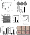
(A) BPDE-transformed WI-26 cells with different expression ratio of AIMP2-DX2 to AIMP2-F were tested for their propensity to anchorage-independent colony formation. The relationship between the ratio of AIMP2-DX2 to AIMP2-F and the average numbers of the produced colonies with standard deviation in each colony were displayed as dot plot. (B) WI-26 cells stably expressing EV, AIMP2-F and -DX2 were also compared for anchorage-independent colony formation. The same numbers of the three cell lines (200 cells per plate) were spread on the testing medium and the resulting colonies were counted and shown as bar graph. The experiments were repeated three times. (C) Two independent 12.5d MEFs were obtained from the wild type and AIMP2-DX2 transgenic mice and compared for the expression levels of p53 and phosphorylation status of p53. Expression of AIMP2-DX2 was also determined by Western blot analysis. Tubulin was used as a loading control. 12.5d MEFs isolated from the wild type and AIMP2-DX2 transgenic mice were treated with adriamycin (1 µM) for 24 h and the sub-G1 populations induced by adriamycin treatment were determined by flow cytometry (D). The cell growth in completed media was also monitored (E). Susceptibility to benzopyrene-induced lung carcinogenesis was compared between the wild type and AIMP2-DX2 transgenic mice by tumor incidence (F) and area (G). The area was represented by the numbers given in Bio-Image J 2.0. (H) Three lungs were isolated from the wild type and AIMP2-DX2 transgenic mice and subjected to hematoxylin and eosin staining.
Figure 7
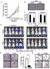
Figure 7. Knock-down of AIMP2-DX2 can retard tumor growth.
(A) NCI-H460 (1×10⁷) cells were injected at both flanks of 6 week old BALB/c nude mice. When volume of tumor mass reached to 250 mm3, anti-DX2 or control siRNAs were intratumorally injected at 50 mg/kg dosage once in three days for four times (n = 7 per each group). The tumor volumes were determined as described in Materials and Methods. (B) Tumors isolated from anti-DX2 and control siRNA treated mice were subjected to immunohistochemistry with the antibodies against p53 (C) Cellular levels of AIMP2-F and –DX2 transcripts in the tissues of the isolated tumors were compared by quantitative RT-PCR. (D) Luciferase-expressing A549 lung cancer cells (1×10⁷) were injected into tail vein of BALB/c nude mice and the dissemination of the cells into lungs was monitored by IVIS (Xenogen). Each group of the injected mice (n = 5/group) intratracheally received si-scramble (50 µg) or si-AIMP2-DX2 (50 µg) mixed with GDM-PEI in 50 µl of 0.9% saline. The tumor growth was monitored by photon flux released from luciferin as described in Materials and Methods. The data were expressed as photon-flux (photons/s/cm2/steradian). The photon flux for each measurement is represented by color scale. (E) Tumor tissues isolated from each group were examined by hetatoxylin and eosin staining. Two examples from each group are displayed. (F) The plasmid DNA encoding shRNA against AIMP2-DX2 was mixed with glucosylated polyethyleneimine (G-PEI), vaporized in humid vacuum chamber and inhaled for 30 minutes through the nose of the mice that contained benzopyrene-induced lung cancers as described in Materials and Methods. The mice were randomly sacrificed at time interval and the tumor growth was determined by the area of tumor tissues in the total measured area (n = 4). (G) Histological analysis of tumor regions by hematoxylin and eosin staining. Arrow heads indicate tumor nodules.
Figure 8
Figure 8. Correlation of expression ratio of AIMP2-DX2 to AIMP2-F with lung cancer stage and survival.
(A) AIMP2-DX2/F ratios of patient tissues with cancer stage IA (n = 5), IB (n = 6) and IIB∼IIIB (n = 7) were compared with that of normal lung tissues (n = 5) by quantitative RT-PCR method as described. The ratios were increased significantly (p<0.05) according to tumor progression. Cancer stages were classified based on TNM (tumor, nodes, metastasis) system. Kaplan-Meier estimates of the (B) OS and (C) DFS according to the level of the expression ratios of AIMP2-DX2 to AIMP2-F in 97 lung cancer patients. The OS was significantly worse in patients with higher expression ratio of AIMP2-DX2 to AIMP2-F than in patients with lower expression ratio. P- values were calculated by the log-rank test.

References

    1. Stetefeld J, Ruegg MA. Structural and functional diversity generated by alternative mRNA splicing. Trends Biochem Sci. 2005;30:515–521. - PubMed
    1. Nissim-Rafinia M, Kerem B. The splicing machinery is a genetic modifier of disease severity. Trends Genet. 2005;21:480–483. - PubMed
    1. Schwerk C, Schulze-Osthoff K. Regulation of apoptosis by alternative pre-mRNA splicing. Mol Cell. 2005;19:1–13. - PubMed
    1. Park SG, Ewalt KL, Kim S. Functional expansion of aminoacyl-tRNA synthetases and their interacting factors: new perspectives on housekeepers. Trends Biochem Sci. 2005;30:569–574. - PubMed
    1. Lee SW, Cho BH, Park SG, Kim S. Aminoacyl-tRNA synthetase complexes: beyond translation. J Cell Sci. 2004;117:3725–3734. - PubMed

Publication types

MeSH terms

Substances