. 2011 Mar 31;6(3):e18475.
doi: 10.1371/journal.pone.0018475.

Comprehensive analysis of transcript start sites in ly49 genes reveals an unexpected relationship with gene function and a lack of upstream promoters

Affiliations

Comprehensive analysis of transcript start sites in ly49 genes reveals an unexpected relationship with gene function and a lack of upstream promoters

Frances Gays et al. PLoS One. .

Abstract

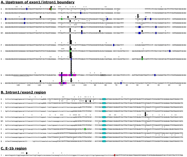
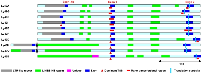
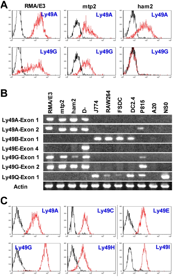
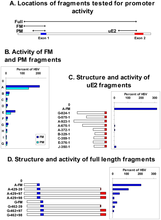
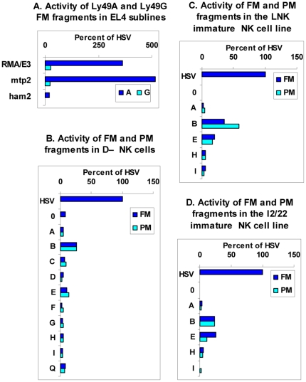
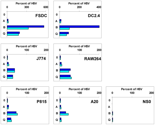
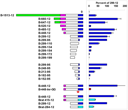
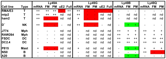
Comprehensive analysis of the transcription start sites of the Ly49 genes of C57BL/6 mice using the oligo-capping 5'-RACE technique revealed that the genes encoding the "missing self" inhibitory receptors, Ly49A, C, G, and I, were transcribed from multiple broad regions in exon 1, in the intron1/exon2 region, and upstream of exon -1b. Ly49E was also transcribed in this manner, and uniquely showed a transcriptional shift from exon1 to exon 2 when NK cells were activated in vitro with IL2. Remarkably, a large proportion of Ly49E transcripts was then initiated from downstream of the translational start codon. By contrast, the genes encoding Ly49B and Q in myeloid cells, the activating Ly49D and H receptors in NK cells, and Ly49F in activated T cells, were predominantly transcribed from a conserved site in a pyrimidine-rich region upstream of exon 1. An ∼200 bp fragment from upstream of the Ly49B start site displayed tissue-specific promoter activity in dendritic cell lines, but the corresponding upstream fragments from all other Ly49 genes lacked detectable tissue-specific promoter activity. In particular, none displayed any significant activity in a newly developed adult NK cell line that expressed multiple Ly49 receptors. Similarly, no promoter activity could be found in fragments upstream of intron1/exon2. Collectively, these findings reveal a previously unrecognized relationship between the pattern of transcription and the expression/function of Ly49 receptors, and indicate that transcription of the Ly49 genes expressed in lymphoid cells is achieved in a manner that does not require classical upstream promoters.


Conflict of interest statement

Competing Interests: The authors have declared that no competing interests exist.

Figures

Figure 1
Figure 1. Transcriptional start sites in Ly49 genes.
The start sites of cap-trapper 5′RACE cDNAs are plotted onto gene sequence alignments of the region upstream of the exon1-intron1 boundary (A), the region surrounding the intron1-exon2 boundary (B), and exon-1b (C). Exonic regions are shown in upper case letters, intronic regions in lower case letters. In the case of the Ly49I exon-1b region, the distinction is artificial as the intronic region ends with a defective splice signal (highlighted in red). Black vertical bars mark sites at which >10% of all transcripts for a given gene originated, the number of bars indicating the actual percentage in units of 10% (eg. 3 bars = 30–40% of all transcripts). Asterisks show other TSSs in these regions. A small number (<5%) of TSSs mapped outside of these egions (see text) and are therefore not shown on the diagram, but are included in the analysis in Table 1. Bases highlighted in blue or green are the TSSs of cDNAs deposited in Genbank, those in green being from the Riken CAP-trapper high efficiency cDNA cloning project. The red T in Ly49A is the TSS for Ly49A in EL4 cells determined by Kubo et al using primer extension and nuclease protection. Regions highlighted in purple in Ly49B and Ly49Q correspond to canonical Inr sequences, the grey region in Ly49B corresponding to a perfectly positioned canonical DRE sequence, and the underlined bases in Ly49B corresponding to GC and TA rich regions with similarity to BRE and TATA sequences . For 9 of the 10 genes, the general disposition of TSSs in fresh cells was not different to that in cultured cells (see Table 1), and the data has therefore been combined. However, for Ly49E the results obtained from fresh cells (Ef) and cultured cells (Ec) were markedly different and are therefore plotted separately.
Figure 2
Figure 2. Structure of the upstream region of Ly49 genes.
The diagram shows the ∼3.5–6 kb region that extends upstream from exon 2 to just beyond the LTR-like repeat sequence that precedes exon -1b (except for Ly49B that lacks a recognizable exon -1b). Exons are shown in dark blue, homologous intronic regions in light blue, LINE/SINE repeats in green, LTR-like repeats in grey, and unique sequence in purple. Dominant TSSs are shown by red triangles, broad transcriptional initiation regions by red bars, and the translational start site by a pale blue line.
Figure 3
Figure 3. Expression of Ly49 genes in various cells.
A. Different sublines of RMA EL4 cells were stained with JR9 anti-Ly49A and 4D11 anti-Ly49G mAbs (red lines) or medium (black lines) followed by AF647-conjugated secondary antibody. B. RT-PCR analysis of Ly49 transcripts in various cell lines. cDNA prepared from equal numbers of cells was amplified using forward primers located in exon 1, exon 2, or exon 4 together with appropriate reverse primers that in combination gave specific amplification of the relevant Ly49. The identity of the Ly49A exon 1 and Ly49G exon 1 amplimers in RMA/E3 cells was confirmed by cloning and sequencing, as was the unexpected presence of Ly49A, G, and Q transcripts in P815 cells, and of Ly49Q transcripts in NS0 cells. C. D NK cells were stained with mAbs against the Ly49s shown (red lines) or with medium (black lines).
Figure 4
Figure 4. Promoter activity of Ly49 gene fragments in EL4 cells.
A. Schematic diagram of the gene fragments tested for promoter activity, namely the PM and FM fragments from the upstream region, the uE2 fragments from the region upstream of exon 2, and the full length ∼2.3 kb fragments that run from the start of the FM fragments to various positions around the start of exon 2. B–D. The RMA/E3 subline of EL4 cells was transfected with pGL4 plasmids containing no insert (0), the HSV promoter, or various Ly49 gene fragments, as follows: B. FM (dark blue bars) and PM (light blue bars) fragments from the upstream region of the Ly49 genes shown. C. The Ly49A FM fragment or fragments of various length extending upstream from the translational start codon in exon 2 of various Ly49 genes. Numbers show the distance of the start and end of each fragment from the start codon, and the diagrams provide a graphical representation of the fragments with intron 1 unshaded and exon 2 in red. D. The FM fragments from the Ly49A and Ly49G genes, or “full length” fragments of various length beginning at the same upstream position as the FM fragments and ending at various positions in exon 2. Numbers show the distance of the start point from the exon1/intron1 boundary, and the distance of the end point from the translational start codon, a negative value meaning upstream and a positive value meaning downstream of the start codon. In the +97 constructs the Ly49 start codon would be in frame with the luciferase start codon, in the +98 constructs it would be out of frame. The diagrams show a graphical representation of the fragments with exon 1 in blue, intron 1 unshaded, and exon 2 in red. In all cases, luciferase activity is expressed as a percentage of that observed with the HSV promoter.
Figure 5
Figure 5. Lack of correlation between promoter activity and endogenous gene expression.
A. The RMA EL4 sublines shown were transfected with pGL4 plasmids containing no insert (not shown), the HSV promoter, or the FM fragments from upstream of the Ly49A (dark blue bars) and Ly49G (light blue bars) genes. B–D. The mature NK cell line D (B), and the immature NK cell lines LNK (C) and I2/22 (D) were transfected with pGL4 plasmids containing no insert (0), or the FM (dark blue bars) and PM (light blue bars) fragments from upstream of the Ly49 genes shown. In all cases, luciferase activity is expressed as a percentage of that observed with the HSV promoter.
Figure 6
Figure 6. Analysis of promoter activity in the upstream region of the Ly49B and Ly49Q genes.
pGL4 plasmids containing no insert (0), the HSV promoter, or the FM (dark blue bars) and PM (light blue bars) fragments from the upstream regions of the Ly49A, B, and Q genes were transfected into the DC lines FSDC and DC2.4, the macrophage lines J774 and RAW264, the mastocytoma P815, and the B cell lines A20 and NS0. Luciferase activity was calculated as a percentage of that observed with the HSV promoter. Note the different scale used for the DC lines.
Figure 7
Figure 7. Mapping of promoter activity in the upstream region of the Ly49B gene.
pGL4 plasmids containing no insert (0), or various fragments from the upstream region of the Ly49B gene were transfected into FSDC cells. Luciferase activity was calculated as a percentage of that observed with the B-299-12 PM fragment. Numbers show the distance of the start and end of each fragment from the exon1/intron1 boundary, and the diagrams provide a graphical representation of the fragments: dark blue, exon 1 (defined as the region downstream of the dominant transcription initiation site); unshaded, region homologous to that found in the same position in other Ly49 genes; purple, region unique to Ly49B (see Figure 1); green, LINE repeat sequence. The B-448-Inr-GG fragment was identical to the B-448-12 fragment but with the two A residues that form the dominant TSSs of the endogenous gene mutated to G residues (shown by a red star). In a separate series of experiments the activity of fragments from the putative rat orthologue of Ly49B, Ly49i8, (light blue bars) was compared to that of the equivalent FM (B-448-12) and PM (B-299-12) fragments of the mouse Ly49B gene. Histograms show the mean values obtained in 3–8 independent experiments with each construct, error bars showing the SEM where this was big enough to display.
Figure 8
Figure 8. Correlation between the promoter activity of transfected fragments and the expression of endogenous Ly49 genes.
The data summarizes the cell lines of various types (T, T cell; NK, NK cell, Mph, macrophage; DC, dendritic cell; Mast, mast cell; B, B cell) that were examined in this study for the expression of Ly49 mRNA transcripts and that were transfected with pGL4-luciferase plasmids containing the various upstream fragments (FM, PM, uE2, and Full) described in detail elsewhere in this manuscript. The level of expression is shown as: ++, high; +, clearly detectable; −, marginal or undetectable; nd, not determined. Green boxes highlight situations where promoter activity was detected in cells that lacked expression of the corresponding endogenous gene. Red boxes highlight situations where promoter activity was marginal or undetectable in cells that expressed the endogenous gene.

Similar articles

Cited by

References

    1. Yokoyama WM, Plougastel BF. Immune functions encoded by the natural killer gene complex. Nat Rev Immunol. 2003;3:304–316. - PubMed
    1. Dimasi N, Biassoni R. Structural and functional aspects of the Ly49 natural killer cell receptors. Immunol Cell Biol. 2005;83:1–8. - PubMed
    1. Raulet DH, Held W, Correa I, Dorfman JR, Wu MF, et al. Specificity, tolerance and developmental regulation of natural killer cells defined by expression of class I-specific Ly49 receptors. Immunol Rev. 1997;155:41–52. - PubMed
    1. Held W, Kunz B. An allele-specific, stochastic gene expression process controls the expression of multiple Ly49 family genes and generates a diverse, MHC-specific NK cell receptor repertoire. Eur J Immunol. 1998;28:2407–2416. - PubMed
    1. Takei F, McQueen KL, Maeda M, Wilhelm BT, Lohwasser S, et al. Ly49 and CD94/NKG2: developmentally regulated expression and evolution. Immunol Rev. 2001;181:90–103. - PubMed

Publication types

MeSH terms

Substances