. 2011 Mar 31;6(3):e18273.
doi: 10.1371/journal.pone.0018273.

Trafficking defect and proteasomal degradation contribute to the phenotype of a novel KCNH2 long QT syndrome mutation

Affiliations

Trafficking defect and proteasomal degradation contribute to the phenotype of a novel KCNH2 long QT syndrome mutation

Anton Mihic et al. PLoS One. .

Abstract

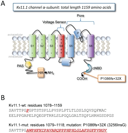
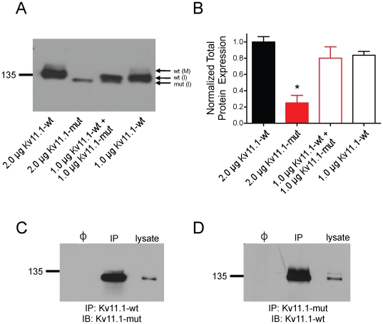
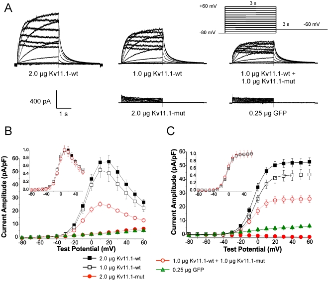
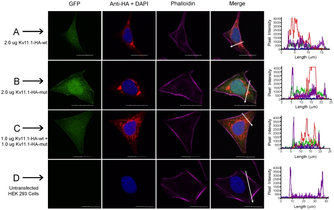
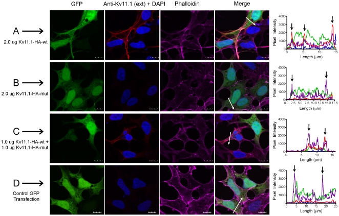
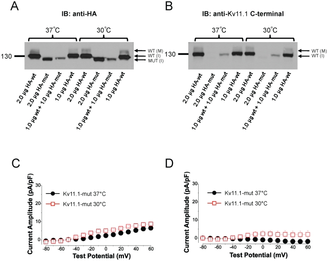
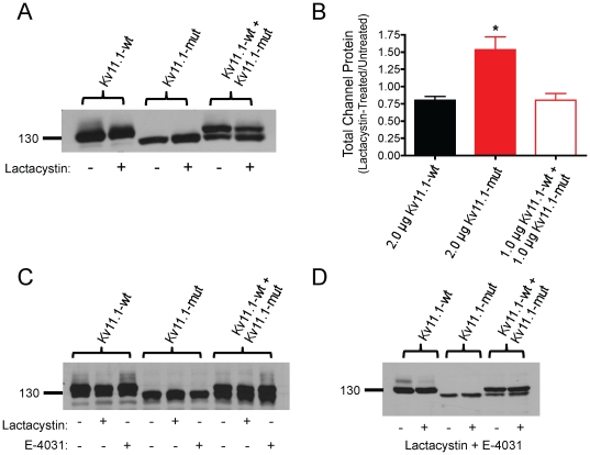
The Kv11.1 (hERG) K+ channel plays a fundamental role in cardiac repolarization. Missense mutations in KCNH2, the gene encoding Kv11.1, cause long QT syndrome (LQTS) and frequently cause channel trafficking-deficiencies. This study characterized the properties of a novel KCNH2 mutation discovered in a LQT2 patient resuscitated from a ventricular fibrillation arrest. Proband genotyping was performed by SSCP and DNA sequencing. The electrophysiological and biochemical properties of the mutant channel were investigated after expression in HEK293 cells. The proband manifested a QTc of 554 ms prior to electrolyte normalization. Mutation analysis revealed an autosomal dominant frameshift mutation at proline 1086 (P1086fs+32X; 3256InsG). Co-immunoprecipitation demonstrated that wild-type Kv11.1 and mutant channels coassemble. Western blot showed that the mutation did not produce mature complex-glycosylated Kv11.1 channels and coexpression resulted in reduced channel maturation. Electrophysiological recordings revealed mutant channel peak currents to be similar to untransfected cells. Co-expression of channels in a 1∶1 ratio demonstrated dominant negative suppression of peak Kv11.1 currents. Immunocytochemistry confirmed that mutant channels were not present at the plasma membrane. Mutant channel trafficking rescue was attempted by incubation at reduced temperature or with the pharmacological agents E-4031. These treatments did not significantly increase peak mutant currents or induce the formation of mature complex-glycosylated channels. The proteasomal inhibitor lactacystin increased the protein levels of the mutant channels demonstrating proteasomal degradation, but failed to induce mutant Kv11.1 protein trafficking. Our study demonstrates a novel dominant-negative Kv11.1 mutation, which results in degraded non-functional channels leading to a LQT2 phenotype.


Conflict of interest statement

Competing Interests: The authors have declared that no competing interests exist.

Figures

Figure 1
Figure 1. 12-lead ECG of proband and sibling.
QTc prolongation was observed before (A) and after (B) electrolyte restoration. C: The asymptomatic sibling carried the same LQT2 mutation and ECG demonstrated mild QTc prolongation with biphasic T waves. D: Epinephrine stress testing was consistent with definite LQTS. Blue arrows denote ST elevation.
Figure 2
Figure 2. Kv11.1 α-subunit schematic and sequence of channel mutation.
A: Located at the C-terminus, the P1086fs+32X (3256InsG) mutation is caused by a guanosine insertion in the codon at position 3256 (2356InsG), which elicits a frameshift at proline 1086 and produces 32 new amino acids before a premature stop codon. The mutation is downstream of the cyclic nucleotide binding domain (cNBD) and produces a truncated channel subunit. The N-terminus contains the Per Arnt Sim domain (PAS) and a HA-tag. B: Sequences for Kv11.1-wt and Kv11.1-mut (P1086fs+32X) including the nonsense 32 amino acid sequence.
Figure 3
Figure 3. Biochemical analysis Kv11.1 protein expression and binding.
A: Immunoblot of equal amounts of protein lysates (25 µg) from HEK cells transfected with 1.0 or 2.0 µg of Kv11.1 cDNA. Kv11.1-wt channels expressed two protein bands corresponding to an immature core-glycosylated 135 kDa ER-resident Kv11.1 protein [wt-(I)], and a mature complex-glycosylated 155 kDa Kv11.1 band [wt-(M)]. Mutant Kv11.1 channels produced a single band at a slightly lower molecular weight (predicted to be 4 kDa smaller than Kv11.1-wt, thus approximately 131 kDa) corresponding to an immature core-glycosylated Kv11.1-mut protein [mut-(I)]. B: Densitometric analysis of total Kv11.1 protein (n = 4 experiments) demonstrated that Kv11.1-mut transfections resulted in significantly less total Kv11.1 protein expression than control or co-transfection (ANOVA *p<0.01). C,D: Reciprocal co-immunoprecipitation of Kv11.1-wt and Kv11.1-mut channels. Cells were transfected with a Kv11.1-wt construct lacking the HA-tag and Kv11.1-HA-mut. Co-immunoprecipitation was performed with anti-Kv11.1-wt antibody (C) (epitope corresponding to C-terminal 16 amino acids) or anti-HA antibody (D) for recognition of Kv11.1-mut. The two channel constructs strongly interacted. (Φ is a sample in which primary antibody was excluded during binding; IP: immunoprecipitation; IB: immunoblot).
Figure 4
Figure 4. Kv11.1 P1086fs+32X mutation reduces peak current amplitude following coexpression with Kv11.1-wt.
Electrophysiological properties of Kv11.1-wt and Kv11.1-mut channels were assessed using whole-cell patch clamping. A: Families of current tracings from −80 to +60 mV following 3 s step depolarizations. Kv11.1-wt currents were reduced following coexpression with Kv11.1-mut, indicating a dominant-negative suppression currents. Kv11.1-mut constructs were indistinguishable from GFP-transfected controls. B: The current-voltage relationship demonstrated that peak current amplitude is significantly reduced following coexpression (2.0 µg Kv11.1-wt, 57.7±4.6 pA/pF, n = 16; 1.0 µg Kv11.1-wt, 51.4±6.3 pA/pF, n = 14; 1.0 µg Kv11.1-wt+1.0 µg Kv11.1-mut, 25.3±2.0 pA/pF, n = 10, p<0.001 from Kv11.1-wt). Peak Kv11.1-mut currents were similar to GFP-transfected cells (2.0 µg Kv11.1-mut, 6.5±0.8 pA/pF, n = 15 versus 0.25 µg GFP, 5.1±0.5 pA/pF, n = 5). The current-voltage profile and C-type inactivation properties were identical following normalization (inset). C: Peak tail currents were measured immediately following repolarization. Kv11.1-wt+Kv11.1-mut tails were significantly reduced compared to control (2.0 µg Kv11.1-wt, 52.8±2.8 pA/pF, n = 16; 1.0 µg Kv11.1-wt, 43.5±3.9 pA/pF, n = 14; 1.0 Kv11.1-wt+1.0 µg Kv11.1-mut, 25.9±2.6 pA/pF, n = 10; p<0.01 from Kv11.1-wt). Tail currents were normalized and fit to a Boltzmann function to assess the steady-state activation properties (inset). No changes in slope or V1/2 parameters were observed.
Figure 5
Figure 5. Detailed analysis of Kv11.1 kinetics.
Channel kinetics were compared between Kv11.1-wt and Kv11.1-wt+Kv11.1-mut groups as no appreciable currents could be measured from Kv11.1-mut alone. There was no difference in channel activation (A), deactivation (B), contribution of the fast component to current decay (C), steady-state inactivation (D), fast inactivation (E) or recovery from inactivation (F).
Figure 6
Figure 6. Total Kv11.1 protein expression in fixed, permeabilized cells.
The staining patterns for cells co-transfected with GFP (green) and HA-tagged Kv11.1 plasmids (CY3, red) were assessed using immunocytochemistry and confocal microscopy. A: Kv11.1-wt; B: Kv11.1-mut; C: co-expression of both plasmids. Untransfected cells served as negative controls (D). DAPI stained nuclei (blue) and phalloidin stained actin filaments (CY5, purple) were used to identify the nucleus and plasma membrane, respectively. White arrows indicate the location of line scans through the plasma membrane and perinuclear regions of merged images. Profile histograms indicate the fluorescence intensity for pixels along line scans for each group. Scale bar represents 20 µm.
Figure 7
Figure 7. Membrane Kv11.1 protein expression in fixed non-permeabilized cells.
Mature Kv11.1 protein expression was investigated using an external Kv11.1 epitope (CY3, red). A: Kv11.1-wt; B: Kv11.1-mut; C: co-expression of Kv11.1-wt and Kv11.1-mut. GFP-transfected cells served as negative controls (D); DAPI stained nuclei (blue); phalloidin stained actin filaments (CY5, purple). White arrows indicate the location of line scans through the plasma membrane and perinuclear regions of merged images. Profile histograms indicate the fluorescence intensity for pixels along line scans for each group. Black arrows indicate the approximate location of plasma membrane in the histogram panels. Scale bar represents 10 µm.
Figure 8
Figure 8. Reduced temperature does not rescue Kv11.1-mut trafficking.
A/B: Cells were incubated at 30°C for 24 h and total Kv11.1 protein was assessed by Western blot. Reduced temperature did not change the intensity of the protein band nor cause the appearance of a Kv11.1-mut mature protein band. Co-transfection of non-HA-tagged Kv11.1-wt and HA-Kv11.1-mut (1.0 µg wt+1.0 µg HA-mut; in lanes 3 and 7) allowed for the specific identification of Kv11.1-mut protein (A; anti-HA antibody) and Kv11.1-wt protein (B; anti-Kv11.1 C-terminal antibody). C: Peak current-voltage relationship for Kv11.1-mut alone at 37°C and 30°C revealed no change in current density (Kv11.1-mut at 37°C, 6.5±0.8 pA/pF, n = 15 versus Kv11.1-mut at 30°C, 8.8±0.9 pA/pF, n = 4). D: Peak tail current amplitude did not significantly change with reduced temperature (Kv11.1-mut at 37°C, −1.8±0.3 pA/pF, n = 15 versus Kv11.1-mut at 30°C, 2.1±2.0 pA/pF).
Figure 9
Figure 9. Trafficking of Kv11.1-mut channels cannot be rescued to the plasma membrane.
A: Incubation with the proteasomal inhibitor lactacystin (20 µM) for 24 h enhanced the expression of immature Kv11.1-mut protein, but did produce a complex-glycosylated Kv11.1-mut protein. B: Densitometric analysis of total protein expression after lactacystin treatment (+) normalized to non-treated lysates (−). There was a significant increase in the expression of total Kv11.1-mut protein compared to the other groups (ANOVA *p<0.01). Untreated Kv11.1-mut cells (2.0 µg Kv11.1-mut, 1.53±0.19, n = 5) versus 2.0 µg Kv11.1-wt control (0.80±0.05) and 1.0 ug Kv11.1-wt+1.0 µg Kv11.1-mut (0.80±0.10, n = 3). C: Twenty-four h treatment with the Kv11.1 channel blocker E-4031 (5 µM) enhanced the mature Kv11.1 protein band in Kv11.1-wt and Kv11.1-wt+Kv11.1-mut groups, but did not elicit a mature Kv11.1-mut channel. D: Combined 24 h treatment with lactacystin (20 µM) and E-4031 (5 µM) did not significantly enhance Kv11.1-mut protein expression, nor did it rescue channel maturation in the Kv11.1-mut or Kv11.1-wt+Kv11.1-mut groups.

References

    1. Sanguinetti MC, Jurkiewicz NK. Two components of cardiac delayed rectifier K+ current. Differential sensitivity to block by class III antiarrhythmic agents. J Gen Physiol. 1990;96:195–215. - PMC - PubMed
    1. Moss AJ. Long QT Syndrome. JAMA. 2003;289:2041–2044. - PubMed
    1. Morita H, Wu J, Zipes DP. The QT syndromes: long and short. Lancet. 2008;372:750–763. - PubMed
    1. Roden DM. Clinical practice. Long-QT syndrome. N Engl J Med. 2008;358:169–176. - PubMed
    1. Anderson CL, Delisle BP, Anson BD, Kilby JA, Will ML, et al. Most LQT2 mutations reduce Kv11.1 (hERG) current by a class 2 (trafficking-deficient) mechanism. Circulation. 2006;113:365–373. - PubMed

Publication types

MeSH terms