. 2011 Mar 31;6(3):e17966.
doi: 10.1371/journal.pone.0017966.

Herpes simplex virus dances with amyloid precursor protein while exiting the cell

Affiliations

Herpes simplex virus dances with amyloid precursor protein while exiting the cell

Shi-Bin Cheng et al. PLoS One. .

Abstract

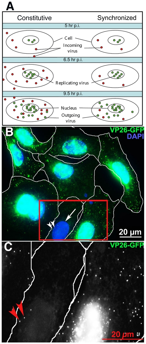
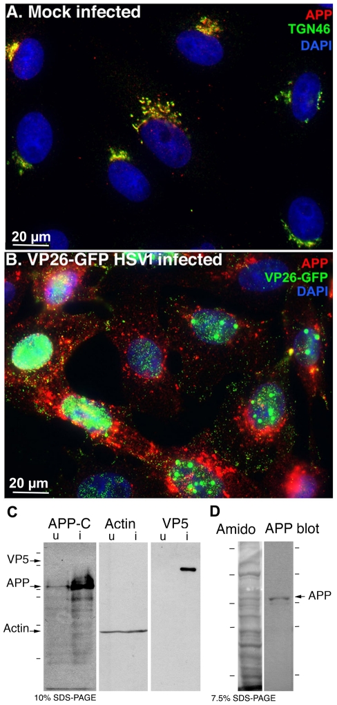
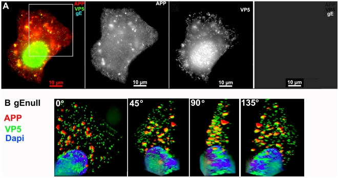
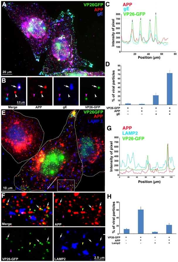
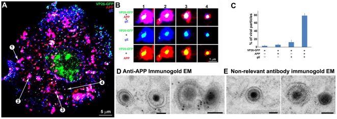
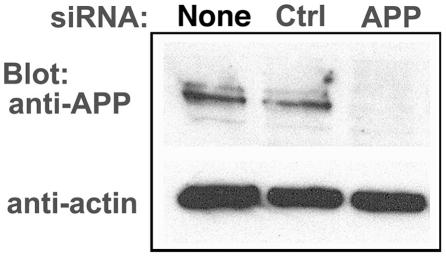
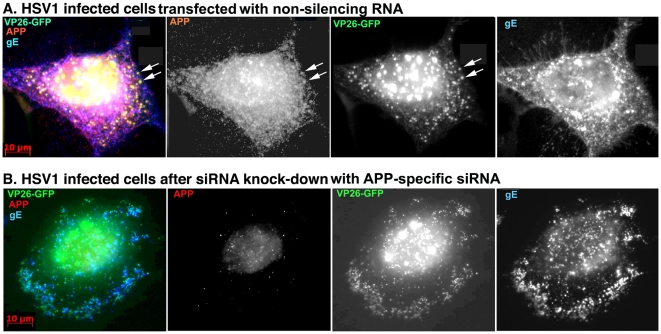
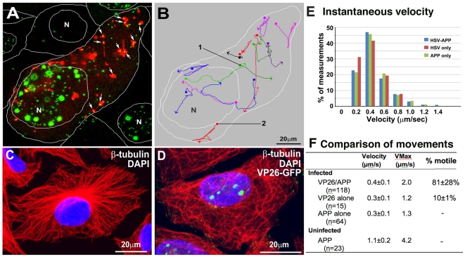
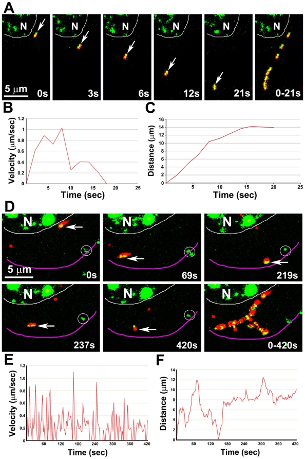
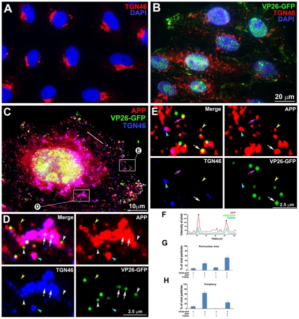
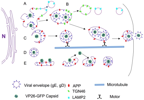
Herpes simplex type 1 (HSV1) replicates in epithelial cells and secondarily enters local sensory neuronal processes, traveling retrograde to the neuronal nucleus to enter latency. Upon reawakening newly synthesized viral particles travel anterograde back to the epithelial cells of the lip, causing the recurrent cold sore. HSV1 co-purifies with amyloid precursor protein (APP), a cellular transmembrane glycoprotein and receptor for anterograde transport machinery that when proteolyzed produces A-beta, the major component of senile plaques. Here we focus on transport inside epithelial cells of newly synthesized virus during its transit to the cell surface. We hypothesize that HSV1 recruits cellular APP during transport. We explore this with quantitative immuno-fluorescence, immuno-gold electron-microscopy and live cell confocal imaging. After synchronous infection most nascent VP26-GFP-labeled viral particles in the cytoplasm co-localize with APP (72.8+/-6.7%) and travel together with APP inside living cells (81.1+/-28.9%). This interaction has functional consequences: HSV1 infection decreases the average velocity of APP particles (from 1.1+/-0.2 to 0.3+/-0.1 µm/s) and results in APP mal-distribution in infected cells, while interplay with APP-particles increases the frequency (from 10% to 81% motile) and velocity (from 0.3+/-0.1 to 0.4+/-0.1 µm/s) of VP26-GFP transport. In cells infected with HSV1 lacking the viral Fc receptor, gE, an envelope glycoprotein also involved in viral axonal transport, APP-capsid interactions are preserved while the distribution and dynamics of dual-label particles differ from wild-type by both immuno-fluorescence and live imaging. Knock-down of APP with siRNA eliminates APP staining, confirming specificity. Our results indicate that most intracellular HSV1 particles undergo frequent dynamic interplay with APP in a manner that facilitates viral transport and interferes with normal APP transport and distribution. Such dynamic interactions between APP and HSV1 suggest a mechanistic basis for the observed clinical relationship between HSV1 seropositivity and risk of Alzheimer's disease.


Conflict of interest statement

Competing Interests: The authors have declared that no competing interests exist.

Figures

Figure 1
Figure 1. Synchronization of viral infection.
(A) Diagram of the "in-coming/out-going" problem. Constitutive infections (left column), in which virus is added to the culture and allowed to remain throughout incubation, results in continuous entry of virus and mixing of in-coming and out-going viral particles in the cytoplasm. In synchronized infection (right column) viral exposure is limited by removal of the media, release of adherent virus from cell surface with acid-glycine and inactivation with human serum, resulting in few in-coming virus in the cytoplasm at later stages. Sub-confluent cultures are required to reduce viral transmission through cell-cell junctions. (B) An example of digital images used to quantify cellular locations of VP26-GFP particles. In this example, cells synchronously infected with VP26-GFP HSV1 (green) were fixed at 7 hr p.i., and counterstained with DAPI (blue). Images were collected by widefield fluorescence to detect VP26-GFP particles throughout the full thickness of the cells. Infected cell nuclei containing VP26-GFP appear turquoise at this exposure. Most of these infected cells display multiple viral particles in the cytoplasm. One cell (arrow) with no nuclear GFP fluorescence has three cytoplasmic GFP-particles (arrowheads) that therefore are in-coming virus. (C) Higher magnification of the boxed region in B in the GFP channel to show absence of diffuse nuclear fluorescence. Red arrows indicate two of the three GFP particles in this cell. See Figure S1 for quantification.
Figure 2
Figure 2. APP relocalizes in infected cells.
(A) APP (red) co-localizes with TGN46 (green) in a compact tuft to one side of the nucleus (DAPI, blue) representing the trans-Golgi network in mock infected cells. (B) In cells infected with VP26-GFP HSV1 (green), APP (red) is distributed in particles throughout the cytoplasm at 7 hr p.i. Nuclei are stained with DAPI (blue). (C) Western blotting of uninfected (u) and infected (i) cells with anti-APP, anti-actin (loading control) and anti-VP5 (viral capsid) demonstrates significant increased amound of APP in infected cells. Actin bands remain similar, and VP5, as expected, is only detected in lanes loaded with infected cell lysate. Note no new APP bands are detected in the infected versus uninfected cell lysates. (D) Isolated virus separated on 7.5% SDS-PAGE and stained with amido black for protein and probed for APP by Western blotting with the same antibodies used for immuno-fluorescence. Note that only the 90–110 kD doublet representing APP is detected by anti-APP, with no additional viral protein bands detected. See also Figure S2 for split channels and Figure S3 for histone staining.
Figure 3
Figure 3. APP co-localizes with gEnull virus.
A representative example of a cell infected with gE null virus stained for APP (red), gE (far red), and VP5 (green), the major capsid protein. (A) Low magnification wide-field image of the three channels, APP, VP5, and gE shown in color with merged channels (left), and then each individual channel, as labeled, of the same image. (B) Higher magnification of the boxed region shown in (A) and rendered in 3D. A Z-stack of 45 focal planes captured at 0.35 µm intervals was deconvolved using iterative processing and rendered in 3D. In four angles of rotation, co-localization of green VP5 capsids and red APP membranes throughout the cytoplasm of a cell infected with gEnull virus is shown. See Movie S1 for a full rotation of the 3D stack shown in (B).
Figure 4
Figure 4. Out-going cytoplasmic VP26-GFP particles co-localize with APP and not with LAMP2.
Cells were synchronously infected with VP26-GFP HSV1 (green) and fixed at 7 hr p.i.. (A–D) Examples of infected cells stained for gE (blue) and APP (red). In (B) a high magnification of boxed region in (A) is shown. A white arrow indicates one of the particles displaying all three labels (gE, APP and VP26). Particles with APP only (white arrowhead) or gE and VP26 (pink arrowhead) are indicated. (F–H) Examples of infected cells stained for LAMP2 (blue) and APP (red). In (F) a high magnification of the boxed regions in (E) is shown. White arrows indicate APP and gE, pink arrowheads indicate LAMP2, and yellow arrowheads indicate APP alone. (C and G) Intensity profiles along a line (white) drawn across the merged image in (A) and (E). Arrows indicate the superposition of peaks for each channel. (D and H) Histograms showing the percentage of VP26-GFP particles in each category. VP26-GFP alone (D: 3.0±1.7% and H: 11.8±3.9%) or with APP (D: 2.0±1.5% and H: 61.3±14.7%); VP26-GFP with gE (D: 21.8±5.5%) or with LAMP2 (H: 4.6±1.6%); and VP26-GFP with both APP and gE (D: 72.8±6.7%), or both APP and LAMP2 (H: 22.3±8.7%). Experiments were performed in triplicate, and 2085 particles in 11 cells were counted in D and 1592 particles in 9 cells in H. See Figure S5 for triple label of gD instead of gE, with APP and VP26-GFP.
Figure 5
Figure 5. Confocal and immuno-gold electron microscopy demonstrate co-localization of both viral (gE) and cellular (APP) membrane proteins with VP26-GFP particles.
(A) Example of a 0.8 µm optical section by confocal imaging of a cell infected with VP26-GFP HSV1 (green), fixed at 8.5 hr p.i., and stained for cellular APP (red) and viral glycoprotein, gE (blue). (B) Galleries of particles showing the co-localization of VP26-GFP with gE and APP. (C) Histogram showing the percentage of VP26-GFP particles in each category. VP26-GFP alone (3.2±2.0%), with APP (5.9±2.4%), with gE (12.4±5.6%) and with both APP and gE (78.4±5.7%). 569 particles in 5 cells were counted. (D) Thin section immunogold electron microscopy of HSV1 infected cells probed with anti-C-APP with protein-A linked 10 nm gold particles. Note single and multiple gold particles decorating membranes surrounding viral capsids in the cytoplasm. Bar = 100 nm. (E) Parallel sections from the same EM block treated with an irrelevant rabbit antibody of similar purity and dilution and probed with protein-A gold. Note the absence of gold labeling of viral particles. Also see Figure S5 for co-localization of VP26-GFP particles with APP and viral protein gD, and Figure S6 for additional immunogold electron micrographs.
Figure 6
Figure 6. APP knock-down by siRNA decreases APP protein >90% by Western blotting.
HSV1 infected cells were transfected in parallel with either vehicle alone (None), non-silencing RNA (Ctrl) or siRNA against APP (APP). After 48 hr cells were scraped into lysis buffer, and loaded in parallel on a 10% gel for electrophoresis followed by transfer to nitrocellulose. The blot was divided in two horizontally, the top half probed for APP and the lower half for actin, a loading control. Non-silencing siRNA has little effect, while siRNA for APP decreases APP band intensity almost entirely, with no significant effect on actin.
Figure 7
Figure 7. Knockdown of APP expression eliminates immuno-fluorescent staining of VP26-GFP particles by anti-APP antibodies.
(A) Representative example of a cell treated with non-silencing control siRNA, infected with VP26-GFP HSV1 (green), fixed at 7 hr p.i. and stained for APP (red), gE (blue). APP staining is bright and diffuse, and co-localizes with VP26-GFP viral particles (arrows). (B) Representative example of a cell treated in parallel to the cell shown in (A) but with siRNA for APP. Note the absence of most APP staining, while gE staining of viral particles remains strong.
Figure 8
Figure 8. Co-transport of VP26-GFP particles and APP-mRFP in live cells.
(A) The first frame of a video sequence captured at 7–9 hr p.i. captured at 3-sec intervals for 900 sec (15 min). Many (64%) VP26-GFP particles (green) co-localize with APP-mRFP compartments (red) in this frame appearing bright yellow (arrows). VP26-GFP particles travel with APP-mRFP vesicles, and sometimes join and separate from APP. White lines show the boundaries between cells and position of the nuclei (N). Also see Movie S2. (B) Tracings of some VP26-GFP particles moving with APP-mRFP. Traces are pseudocolored for ease of visualization. Dots and arrowheads indicate the beginning and end of movements, respectively. (C–D) Microtubules are in disarray in HSV1-infected cells. (C) Example of mock-infected and (D) VP26-GFP HSV1 infected cells stained for β-tubulin (red) and DAPI (blue) at 7–9 hr p.i. and imaged by laser scanning confocal. See also Figure S7. (E) Histogram of the instantaneous velocities in infected cells of dual VP26-GFP-APP, solo VP26-GFP and solo APP only particles. Instantaneous velocities of VP26-GFP-APP (n = 118), solo VP26-GFP (n = 15) and solo APP (n = 64) particles in 20 cells in 6 experiments were measured. (F) Table of movements. Quantitative comparison of average instantaneous velocity, maximum velocity and frequency of moves (% motile) for each type of particle from 3–5 different cells from 3 independent experiments, where n =  number particles measured. Note the decreased frequency of movements of the VP26-GFP particles without APP-mRFP, third column. See also Movie S2, Figure S7 and S8, and Table S1.
Figure 9
Figure 9. Representative movements of dual APP-mRFP and VP26-GFP particles.
(A) A double labeled GFP-mRFP particle moves away from the nucleus (arrow, from Movie S2 shown in Figure 5B, particle 1). The last panel shows 8 frames superimposed to demonstrate the pathway. See Movie S3. (B, C) Plots of velocity (B) and distance (C) versus time of particle 1 (in A). (D) A GFP-mRFP particle (arrow, from the video shown in Figure 5B, particle 2) moving away from the nucleus and ending at the periphery of the cell. Last panel shows 23 frames from a time-lapse sequence captured at 3-sec intervals (selected from a total of 141 frames of a 420 sec video) superimposed to demonstrate the pathway. The particle moves back and forth and along multiple tracks, and changes its shape during the movement. Circles indicate stationary GFP particles lacking APP-mRFP. See Movie S4. (E, F) Plots of velocity (E) and distance (F) versus time of particle 2 (in D), show the properties of the movements of each particle. Instantaneous velocity varies widely, with fast and slow velocities alternating.
Figure 10
Figure 10. APP and TGN46 do not co-localize with VP26-GFP labeled viral particles in the periphery of HSV1 infected cells: Evidence for specific APP-HSV1 interactions.
(A) Uninfected cells stained for TGN46 (red) show compact tufts on one side of the DAPI-stained nuclei (blue). (B) Cells synchronously infected with VP26-GFP HSV1 (green), fixed and stained for TGN46 (red) and DAPI for nuclei (blue). (C) Higher magnification of an example of cell infected with VP26-GFP-HSV1 (green) fixed at 7 h p.i. and stained for TGN46 (blue) and APP (red). (D) Co-localization in the peri-nuclear region shown at high magnification of boxed region "D" in cell shown in (C). Arrows indicate co-localization of VP26-GFP particles with APP in a TGN46-stained compartment. White arrowhead indicates a VP26-GFP particle apparently on the surface of a TGN46-stained vesicle. Yellow and cyan arrowheads indicate examples of single anti-APP-stained and both APP and TGN46 stained particles, respectively. (E) Loss of co-localization at the periphery is shown at high magnification of boxed region E at the periphery of the cell shown in (C). Pink and white arrows indicate co-localization of VP26-GFP HSV1 with APP alone at the periphery and in the cytoplasm close to the periphery, respectively. Yellow and cyan arrowheads indicate examples of single APP and both APP and TGN46 labels, respectively. (F) Linescan intensity profile of a region in the intermediate cytoplasm as seen in (C) shows both coincident (arrows) and non-coincident (arrowheads) peaks of TGN46 staining (blue line) with APP (red) and VP26-GFP particles (green). (G) Histogram of particles in the peri-nuclear region showing the percentage of VP26-GFP particles that co-localized with APP and TGN46. The majority of VP26-GFP particles co-localized with APP and TGN46 (51.8±9.5%) and fewer co-localized only with TGN46 without APP (11.6±3.1%). (H) Histogram of particles in the periphery showing the percentage of VP26-GFP particles that co-localized with APP and TGN46. None co-localized with TGN46 alone, although 25.5±14.2% were co-localized with both APP and TGN46. Note that many fewer particles co-localized with TGN46 in the periphery than in the peri-nuclear region, suggesting that membrane compartments co-localized with viral products retain the ability to sort their components. N = 10 cells, 3,782 particles from three experiments.
Figure 11
Figure 11. Diagram.
A cartoon showing various types of interactions between cellular APP and VP26-GFP labeled viral particles documented here. (A) In the peri-nuclear region, VP26-GFP particles dance around and within large peri-nuclear compartments co-localizedd with viral envelope proteins, gE and gD, and cellular membrane proteins, LAMP2, TGN46 and APP. LAMP2 compartments separate from this apparent Golgi network early and rarely co-localize with viral components at the periphery. Some membrane systems with VP26-GFP also label for both APP and TGN46, primarily near the nucleus at the time points studied here. (B) TGN46 particles separate from VP26-GFP labeled viral components farther towards the periphery, while the APP particles remain with VP26-GFP particles and with viral envelope glycoproteins, gE and gD, en route towards the cell surface. (C) VP26-GFP particles may enter smaller post-Golgi APP-staining particles that undergo directed transport. (D) Some VP26-GFP particles remain separate from APP after leaving the nucleus. These may be inside unlabelled membrane systems or be free in the cytoplasm, some have the capacity to transport without APP. (E) VP26-GFP particles may ride on the cytoplasmic surface of APP-labeled membrane systems, come on or off these membranes, or bud into them. Any particular viral particle may employ all of these mechanisms during transit in the cytoplasm. In each case, we hypothesize that microtubule motors, such as kinesin, are recruited, possibly via APP or another cellular motor receptor.

References

    1. Lycke E, Kristensson K, Svennerholm B, Vahlne A, Ziegler R. Uptake and transport of herpes simplex virus in neurites of rat dorsal root ganglia cells in culture. J Gen Virol. 1984;65(Pt 1):55–64. - PubMed
    1. Kristensson K, Lycke E, Roytta M, Svennerholm B, Vahlne A. Neuritic transport of herpes simplex virus in rat sensory neurons in vitro. Effects of substances interacting with microtubular function and axonal flow [nocodazole, taxol and erythro-9-3-(2-hydroxynonyl)adenine]. J Gen Virol. 1986;67(Pt 9):2023–2028. - PubMed
    1. Enquist LW, Tomishima MJ, Gross S, Smith GA. Directional spread of an alpha-herpesvirus in the nervous system. Vet Microbiol. 2002;86:5–16. - PubMed
    1. Holland DJ, Miranda-Saksena M, Boadle RA, Armati P, Cunningham AL. Anterograde transport of herpes simplex virus proteins in axons of peripheral human fetal neurons: an immunoelectron microscopy study. J Virol. 1999;73:8503–8511. - PMC - PubMed
    1. Miranda-Saksena M, Armati P, Boadle RA, Holland DJ, Cunningham AL. Anterograde transport of herpes simplex virus type 1 in cultured, dissociated human and rat dorsal root ganglion neurons. J Virol. 2000;74:1827–1839. - PMC - PubMed

Publication types

MeSH terms

Substances