. 2011 Mar 31;6(3):e18036.
doi: 10.1371/journal.pone.0018036.

Structuring detergents for extracting and stabilizing functional membrane proteins

Affiliations

Structuring detergents for extracting and stabilizing functional membrane proteins

Rima Matar-Merheb et al. PLoS One. .

Abstract

Background: Membrane proteins are privileged pharmaceutical targets for which the development of structure-based drug design is challenging. One underlying reason is the fact that detergents do not stabilize membrane domains as efficiently as natural lipids in membranes, often leading to a partial to complete loss of activity/stability during protein extraction and purification and preventing crystallization in an active conformation.

Methodology/principal findings: Anionic calix[4]arene based detergents (C4Cn, n=1-12) were designed to structure the membrane domains through hydrophobic interactions and a network of salt bridges with the basic residues found at the cytosol-membrane interface of membrane proteins. These compounds behave as surfactants, forming micelles of 5-24 nm, with the critical micellar concentration (CMC) being as expected sensitive to pH ranging from 0.05 to 1.5 mM. Both by 1H NMR titration and Surface Tension titration experiments, the interaction of these molecules with the basic amino acids was confirmed. They extract membrane proteins from different origins behaving as mild detergents, leading to partial extraction in some cases. They also retain protein functionality, as shown for BmrA (Bacillus multidrug resistance ATP protein), a membrane multidrug-transporting ATPase, which is particularly sensitive to detergent extraction. These new detergents allow BmrA to bind daunorubicin with a Kd of 12 µM, a value similar to that observed after purification using dodecyl maltoside (DDM). They preserve the ATPase activity of BmrA (which resets the protein to its initial state after drug efflux) much more efficiently than SDS (sodium dodecyl sulphate), FC12 (Foscholine 12) or DDM. They also maintain in a functional state the C4Cn-extracted protein upon detergent exchange with FC12. Finally, they promote 3D-crystallization of the membrane protein.

Conclusion/significance: These compounds seem promising to extract in a functional state membrane proteins obeying the positive inside rule. In that context, they may contribute to the membrane protein crystallization field.


Conflict of interest statement

Competing Interests: The authors have declared that no competing interests exist.

Figures

Figure 1
Figure 1. Concept of salt bridge network between anionic and amphiphilic molecules and basic residues located at the cytosol-membrane interface of membrane proteins.
(A) Scheme of a hypothetical dimeric membrane protein typically displaying basic residues at the cytosol-membrane interface, as established by von Heijne . In the absence of lipids, the membrane domain remains in a native conformation due to compounds displaying detergent properties for keeping the membrane protein in solution (grey molecules), but also mild-anionic groups (black molecules), for generating a network of salt bridges close to the membrane domain with basic amino acids carried by the intracellular loops (or domains) of the membrane proteins. (B) Chemical structure of the designed molecules, C4Cn. Three aromatic rings are substituted by a methylene carboxyl group, -CH2COOH, at the para position. An aliphatic chain R, O(CH2)0-11CH3, is grafted onto the fourth phenolic group. The resulting 3D-structure is modelled from the crystal structure of nitrile derivatives , substituting the CN groups with carboxyl groups.
Figure 2
Figure 2. C4Cn behaviour in solution and interaction with basic amino acids.
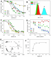
(A) Effects of increasing C4C1, C4C3, C4C7 and C4C12 concentrations on the surface tension of aqueous solution. The surface tension is measured as described in Methods. Each value is the mean of three experiments ± the standard error. C4C1, C4C3, C4C7 and C4C12 are indicated by circles, diamonds, triangles and squares, respectively. (B) Dynamic light scattering of C4Cn in aqueous solution. Experiments were carried out as described in Methods. Mean size values are indicated in nm for the corresponding compound. (C) Effect of pH on the surface tension generated by C4C7. Experiments have been carried out as in (A), neutralizing C4C7 at pH 9.0, 8.0 and 6.0, and measuring the resulting surface tension by increasing the concentration of the compound, as indicated by circles, triangles and squares, respectively. The values result from triplicate experiments. (D) Interaction of C4C7 with amino acids probed by surface tension. C4C7 diluted to 10 µM was incubated with increasing concentrations of either glutamate (circles), lysine (triangles), arginine (squares) or proline (diamonds), measuring the resulting surface tension, as described in Methods. (E) Chemical shifts of the αH and εH protons of lysine in the presence of C4C1. The 1H NMR spectra of L-Lysine (10 mM) was recorded as described in Methods in the presence of increasing C4C1 concentrations as indicated in the Figure, resulting in chemical shifts of αH and εH protons of lysine which were plotted as a function of C4C1 concentration. (F). Dissociation of the L-lysine - C4C1 complex, each added at 10 mM, induced by increasing the salt concentration as indicated and probed by measuring the chemical shift of lysine αH protons.
Figure 3
Figure 3. Extraction of membrane proteins by C4Cn derivatives.
Extraction of ABC transporters by C4Cn, prokaryotic BmrA (A) and YheI/YheH (B) expressed in E. coli, human ABCG2 expressed in Sf9 (C) or HEK293 (D) cells, together with AcrB expressed in E. coli (E) and the SR-Ca2+-ATPase (F), was carried out on the corresponding membrane fractions as described in Methods. After solubilisation, extracted and non-extracted materials were separated by high-speed centrifugation, generating a supernatant S and a pellet P which were loaded on a 10% SDS-PAGE, and after migration either stained with Coomassie blue or submitted to a Western blot for ABCG2 (upper lanes in panels in C and D). Arrows indicate the position of each monomer. Positive control experiments were carried out with DDM, FC12 and C12E8, negative controls were carried out without detergent (No det). The red dotted line indicates the threshold of extraction.
Figure 4
Figure 4. BmrA purification with C4Cn and detergent exchange.
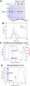
(A) SDS-PAGE of the sequential extraction of BmrA. As detailed in Methods, the membrane fraction (lane T) was incubated with C4C3 and then centrifuged to give the supernatant S and the pellet P. The latter, enriched in BmrA, was suspended in the presence of C4C7 and then centrifuged to give the corresponding supernatant S and pellet P. Arrows indicate the position of BmrA, C4C3 and C4C7. The C4C7 supernatant was then subjected to DLS (B), Ni-affinity chromatography (C) and gel filtration carried out with FC12 (D) from which respective pools indicated by stars were loaded onto SDS-PAGE.
Figure 5
Figure 5. C4Cn preserve a functional state of BmrA.
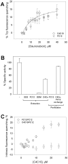
(A) Binding of daunorubicin to BmrA monitored by intrinsic (Tryptophan, Trp) fluorescence quenching. BmrA was extracted and purified either with C4C10 (circles) or FC12 (triangles) as in Figure 4, the former being subsequently exchanged by FC12 as in Figure 4D. The purified protein was then incubated with increasing concentrations of daunorubicin, the binding of the drug being probed by the variation of intrinsic fluorescence of BmrA, as described in Methods. (B) VO4-sensitive ATPase activity of BmrA (see Methods) in different fractions: BmrA-enriched membrane fraction (“-“ bar) corresponding to 0.5 µmol/min.mg and taken as 100%; BmrA-enriched membrane fraction solubilized with 1% SDS, FC12, DDM, or C4C3+C4C7 (“Extraction/C4Cn” bar, carried out as in Fig. 4); BmrA extracted with FC12 and then purified by metal affinity and gel filtration with FC12 (“Purification/FC12” bar); BmrA extracted with C4C3+C4C7 and then purified by metal affinity with C4C7 followed by detergent exchange with FC12 using gel filtration as carried out in Figure 4 (“Purification/C4Cn, FC12 exchange” bar). (C) Intrinsic fluorescence quenching monitoring of C4C10 binding on BmrA. BmrA was extracted either with C4C10 or FC12 and then purified with FC12 as in Figure 4 generating two populations on which C4C10 binding was monitored by probing the quenching of intrinsic fluorescence of 1 µM BmrA as detailed in Methods.
Figure 6
Figure 6. Crystallization of BmrA extracted with C4C10.
BmrA was extracted and purified as described in Figure 5, using C4C10 instead of C4C7 and exchanging it with FC12. (A) The protein, concentrated to 10 mg/ml, and mixed with 1 mM doxorubicin, crystallized after 10 days in 0.2 M KSCN, 20% PEG 3350, and was (B) analyzed at the ESRF beamline ID23EH-2.

References

    1. Wallin E, Von-Heijne G. Genome-wide analysis of integral membrane proteins from eubacterial, archaean, and eukaryotic organisms. Protein Sci. 1998;7:1029–1038. - PMC - PubMed
    1. Overington JP, Al-Lazikani B, Hopkins AL. How many drug targets are there? Nat Rev Drug Discov. 2006;5:993–996. - PubMed
    1. White SH. Membrane Proteins of known 3D structure. 2009 Available: http://blanco.biomol.uci.edu/Membrane_Proteins_xtal.html. Accessed 2011 March 11.
    1. Seddon AM, Curnow P, Booth PJ. Membrane proteins, lipids and detergents: not just a soap opera. Biochim Biophys Acta. 2004;1666:105–117. - PubMed
    1. van Meer G, Voelker DR, Feigenson GW. Membrane lipids: where they are and how they behave. Nat Rev Mol Cell Biol. 2008;9:112–124. - PMC - PubMed

Publication types

MeSH terms