. 2011 Jun;7(2):231-41.
doi: 10.1007/s11302-011-9228-9. Epub 2011 Apr 2.

Impact of CD39 and purinergic signalling on the growth and metastasis of colorectal cancer

Affiliations

Impact of CD39 and purinergic signalling on the growth and metastasis of colorectal cancer

Beat M Künzli et al. Purinergic Signal. 2011 Jun.

Abstract

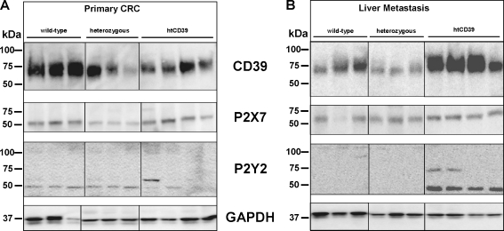
Despite improvements in prevention and management of colorectal cancer (CRC), uncontrolled tumor growth with metastatic spread to distant organs remains an important clinical concern. Genetic deletion of CD39, the dominant vascular and immune cell ectonucleotidase, has been shown to delay tumor growth and blunt angiogenesis in mouse models of melanoma, lung and colonic malignancy. Here, we tested the influence of CD39 on CRC tumor progression and metastasis by investigating orthotopic transplanted and metastatic cancer models in wild-type BALB/c, human CD39 transgenic and CD39 deficient mice. We also investigated CD39 and P2 receptor expression patterns in human CRC biopsies. Murine CD39 was expressed by endothelium, stromal and mononuclear cells infiltrating the experimental MC-26 tumors. In the primary CRC model, volumes of tumors in the subserosa of the colon and/or rectum did not differ amongst the treatment groups at day 10, albeit these tumors rarely metastasized to the liver. In the dissemination model, MC-26 cell line-derived hepatic metastases grew significantly faster in CD39 over-expressing transgenics, when compared to CD39 deficient mice. Murine P2Y2 was significantly elevated at both mRNA and protein levels, within the larger liver metastases obtained from CD39 transgenic mice where changes in P2X7 levels were also noted. In clinical samples, lower levels of CD39 mRNA in malignant CRC tissues appeared associated with longer duration of survival and could be linked to less invasive tumors. The modulatory effects of CD39 on tumor dissemination and differential levels of CD39, P2Y2 and P2X7 expression in tumors suggest involvement of purinergic signalling in these processes. Our studies also suggest potential roles for purinergic-based therapies in clinical CRC.


Figures

Fig. 1
Fig. 1
Experimental assessments of tumor growth and dissemination. a Representative tumor growth of primary MC-26 orthotopic transplant cancer tumor formation in the colorectum. The dotted lines indicate the tumor border. b Liver metastases grew significantly larger in human CD39 transgenic mouse livers, compared to heterozygous animals after intrasplenic injections. c In the colorectal orthotopic model, no differences in the three-dimensional tumor bulk were calculated amongst the groups. d In the dissemination liver metastases model, significant differences in liver weights (due to metastatic growth) were observed, comparing livers from hz and htCD39 livers (P < 0.05). e In the orthotopic colorectal cancer model, macroscopic liver metastases could not be detected within the three groups. Microscopic metastases were excluded by histological assessment (not shown). f Expression patterns of CD39, CD39L1, P2X7 and P2Y2 mRNA were determined. MC-26 colorectal cancer cells do not express CD39
Fig. 2
Fig. 2
Immunopathology of CD39 in CRC. a Immunohistochemical staining for mouse CD39 revealed expression in peritumoral stroma cells, endothelial and mononuclear cells but not in MC-26 tumor cells in orthotopic tumors. b The tumor border in orthotopic tumors is strongly CD39 positive, comprising mononuclear, stromal and endothelial cells. c Invading tumor clusters of MC-26 cells show strong staining for CD39 at the highly proliferative endothelial and stromal cells at the tumor border. df In liver metastases, specific staining for CD39 was displayed by hepatic fibroblasts, endothelial and mononuclear cells. The formation of spreading ductal cancer clusters (metastases) was only observed in d wild-type and e heterozygous livers. f Large solid CRC metastases that comprised the majority of the liver tissue sections were observed in htCD39 liver tissue slides
Fig. 3
Fig. 3
CD39 and P2 receptors in CRC. a In orthotopic tumors, CD39, P2X7 and P2Y2 mRNA appeared at comparable levels to those seen in control livers. b As expected, mouse CD39 mRNA was significantly lower expressed in genetically deficient over wild-type and htCD39 tissues. P2X7 mRNA was significantly over-expressed in htCD39, compared to wt and deficient, heterozygous tissues. P2Y2 mRNA expression was significantly upregulated in the more substantive htCD39 liver metastases, whereas P2Y2 mRNA was expressed at low levels in wt and hz tissues. c Significant decreases in human CD39 mRNA expression occurred in T3N + M0 (18–36 months), T3N + M1 hep (synchron) and T4N0M0 tumors, when compared to control tissues. d Significantly less expression of P2X7 mRNA was observed in T3N + M0 (72–110 months) and T3N + M1 hep (metachron) tumors vs. normal colon. e P2Y2 mRNA expression was significantly lower in T3N + M1 hep (synchron) and T3N + M1 hep (metachron) colorectal cancer specimens, compared to normal colon
Fig. 4
Fig. 4
Western blot analysis of CRC. a In primary colorectal cancer, mouse CD39 was lower in heterozygous (hz) and htCD39 CRC cells compared to wild-type (wt). P2X7 expression was decreased in CRC tumors in CD39 deficient mice. No changes in protein expression were noted for P2Y2. b Distinct and significant increases of mouse CD39 was observed in liver metastases of CRC in htCD39 mice. P2X7 was slightly over-expressed in htCD39 liver metastases only. P2Y2 mRNA was significantly over-expressed in htCD39 mice, compared to wt and heterozygous samples
Fig. 5
Fig. 5
Purinergic studies in human CRC. Protein expression levels of CD39, P2X7 and P2Y2 (ac) and localization of P2Y2 (d, e) was determined in human CRC. Changes in protein levels were observed for P2Y2 (c), which was significantly less in T3N + M0 (72–110 months; P < 0.01) and T3N + M1 hep (metachron; P < 0.05), respectively over controls. No such changes were noted for CD39 (a) and P2X7 (b). P2Y2 expression was confirmed in human normal tissue (d) and colorectal cancer (e) to be localized to mononuclear cells, stromal cells such as fibroblasts and on nerves (e) but was not noted on normal or cancerous epithelial cells

References

    1. Brenner H, Stegmaier C, Ziegler H. Long-term survival of cancer patients in Germany achieved by the beginning of the third millenium. Ann Oncol. 2005;16(6):981–986. doi: 10.1093/annonc/mdi186. - DOI - PubMed
    1. Takahashi T, Morotomi M, Nomoto K. A novel mouse model of rectal cancer established by orthotopic implantation of colon cancer cells. Cancer Sci. 2004;95(6):514–519. doi: 10.1111/j.1349-7006.2004.tb03242.x. - DOI - PMC - PubMed
    1. Kramer I, Lipp HP. Bevacizumab, a humanized anti-angiogenic monoclonal antibody for the treatment of colorectal cancer. J Clin Pharm Ther. 2007;32(1):1–14. doi: 10.1111/j.1365-2710.2007.00800.x. - DOI - PubMed
    1. Bresalier RS, Hujanen ES, Raper SE, Roll FJ, Itzkowitz SH, Martin GR, Kim YS. An animal model for colon cancer metastasis: establishment and characterization of murine cell lines with enhanced liver-metastasizing ability. Cancer Res. 1987;47(5):1398–1406. - PubMed
    1. Morikawa K, Walker SM, Nakajima M, Pathak S, Jessup JM, Fidler IJ. Influence of organ environment on the growth, selection, and metastasis of human colon carcinoma cells in nude mice. Cancer Res. 1988;48(23):6863–6871. - PubMed