

The role of CCN2 in cartilage and bone development
- PMID: 21484188
- PMCID: PMC3145877
- DOI: 10.1007/s12079-011-0123-5
The role of CCN2 in cartilage and bone development
Abstract
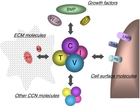
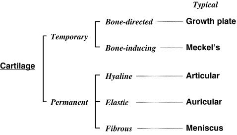
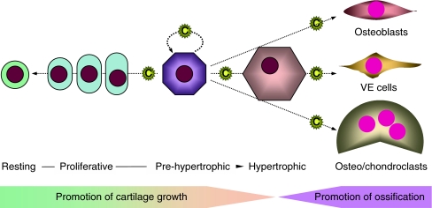
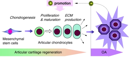
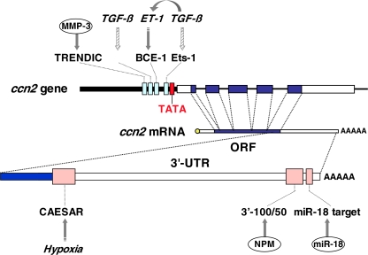
CCN2, a classical member of the CCN family of matricellular proteins, is a key molecule that conducts cartilage development in a harmonized manner through novel molecular actions. During vertebrate development, all cartilage is primarily formed by a process of mesenchymal condensation, while CCN2 is induced to promote this process. Afterwards, cartilage develops into several subtypes with different fates and missions, in which CCN2 plays its proper roles according to the corresponding microenvironments. The history of CCN2 in cartilage and bone began with its re-discovery in the growth cartilage in long bones, which determines the skeletal size through the process of endochondral ossification. CCN2 promotes physiological developmental processes not only in the growth cartilage but also in the other types of cartilages, i.e., Meckel's cartilage representing temporary cartilage without autocalcification, articular cartilage representing hyaline cartilage with physical stiffness, and auricular cartilage representing elastic cartilage. Together with its significant role in intramembranous ossification, CCN2 is regarded as a conductor of skeletogenesis. During cartilage development, the CCN2 gene is dynamically regulated to yield stage-specific production of CCN2 proteins at both transcriptional and post-transcriptional levels. New functional aspects of known biomolecules have been uncovered during the course of investigating these regulatory systems in chondrocytes. Since CCN2 promotes integrated regeneration as well as generation (=development) of these tissues, its utility in regenerative therapy targeting chondrocytes and osteoblasts is indicated, as has already been supported by experimental evidence obtained in vivo.
Figures
Similar articles
-
CCN2: a master regulator of the genesis of bone and cartilage.J Cell Commun Signal. 2013 Aug;7(3):191-201. doi: 10.1007/s12079-013-0204-8. J Cell Commun Signal. 2013. PMID: 23794334 Free PMC article.
-
Role of CCN2/CTGF/Hcs24 in bone growth.Int Rev Cytol. 2007;257:1-41. doi: 10.1016/S0074-7696(07)57001-4. Int Rev Cytol. 2007. PMID: 17280894 Review.
-
CCN family 2/connective tissue growth factor (CCN2/CTGF) stimulates proliferation and differentiation of auricular chondrocytes.Osteoarthritis Cartilage. 2008 Jul;16(7):787-95. doi: 10.1016/j.joca.2007.11.001. Epub 2008 Mar 4. Osteoarthritis Cartilage. 2008. PMID: 18289887
-
The regenerative effects of CCN2 independent modules on chondrocytes in vitro and osteoarthritis models in vivo.Bone. 2014 Feb;59:180-8. doi: 10.1016/j.bone.2013.11.010. Epub 2013 Nov 20. Bone. 2014. PMID: 24269276
-
[CCN family genes in the development and differentiation of cartilage tissues].Clin Calcium. 2006 Mar;16(3):486-92. Clin Calcium. 2006. PMID: 16508133 Review. Japanese.
Cited by
-
Deer antler extract potentially facilitates xiphoid cartilage growth and regeneration and prevents inflammatory susceptibility by regulating multiple functional genes.J Orthop Surg Res. 2021 Mar 22;16(1):208. doi: 10.1186/s13018-021-02350-4. J Orthop Surg Res. 2021. PMID: 33752715 Free PMC article.
-
Regulation of CCN1 via the 3'-untranslated region.J Cell Commun Signal. 2013 Aug;7(3):207-17. doi: 10.1007/s12079-013-0202-x. Epub 2013 May 16. J Cell Commun Signal. 2013. PMID: 23677691 Free PMC article.
-
New Functions of Classical Compounds against Orofacial Inflammatory Lesions.Medicines (Basel). 2018 Nov 1;5(4):118. doi: 10.3390/medicines5040118. Medicines (Basel). 2018. PMID: 30388792 Free PMC article. Review.
-
Genome-wide DNA methylation analysis of articular chondrocytes identifies TRAF1, CTGF, and CX3CL1 genes as hypomethylated in osteoarthritis.Clin Rheumatol. 2017 Oct;36(10):2335-2342. doi: 10.1007/s10067-017-3667-9. Epub 2017 May 3. Clin Rheumatol. 2017. PMID: 28470428
-
OstemiR: a novel panel of microRNA biomarkers in osteoblastic and osteocytic differentiation from mesencymal stem cells.PLoS One. 2013;8(3):e58796. doi: 10.1371/journal.pone.0058796. Epub 2013 Mar 22. PLoS One. 2013. PMID: 23533592 Free PMC article.
References
LinkOut - more resources
Full Text Sources
Miscellaneous
