. 2011 Jul;31(5):765-73.
doi: 10.1007/s10571-011-9682-5. Epub 2011 Apr 12.

Intraperitoneal aminoguanidine improves sciatic nerve ischemia-reperfusion injury in male sprague-dawley rats

Affiliations

Intraperitoneal aminoguanidine improves sciatic nerve ischemia-reperfusion injury in male sprague-dawley rats

Mohsen Alipour et al. Cell Mol Neurobiol. 2011 Jul.

Abstract

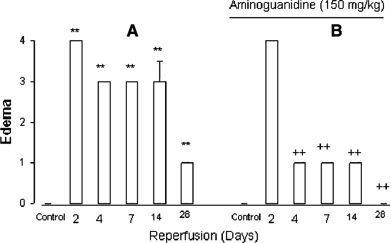
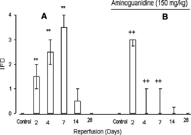
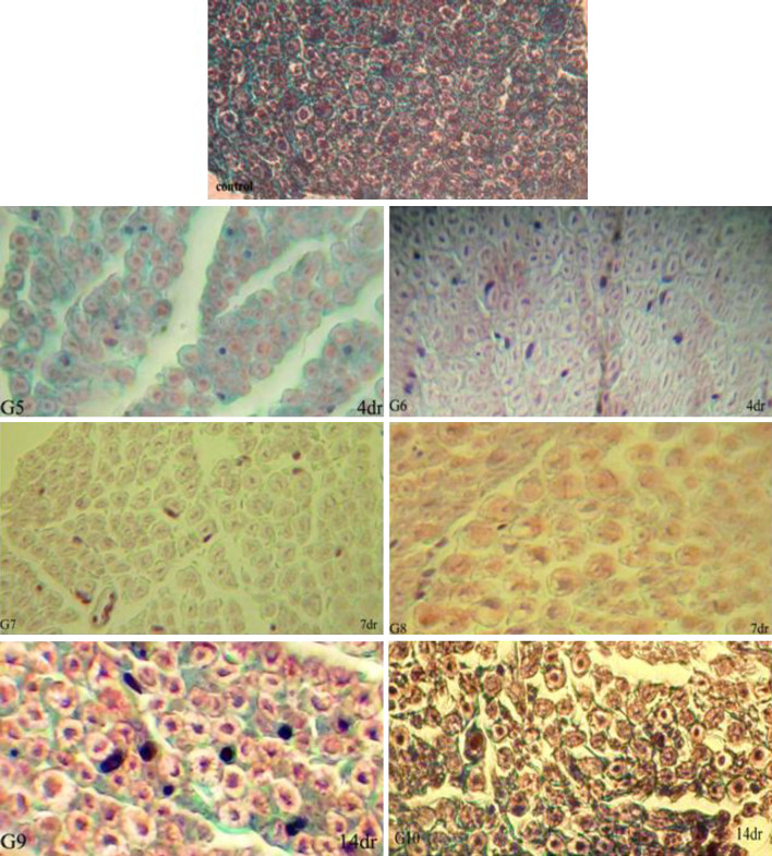
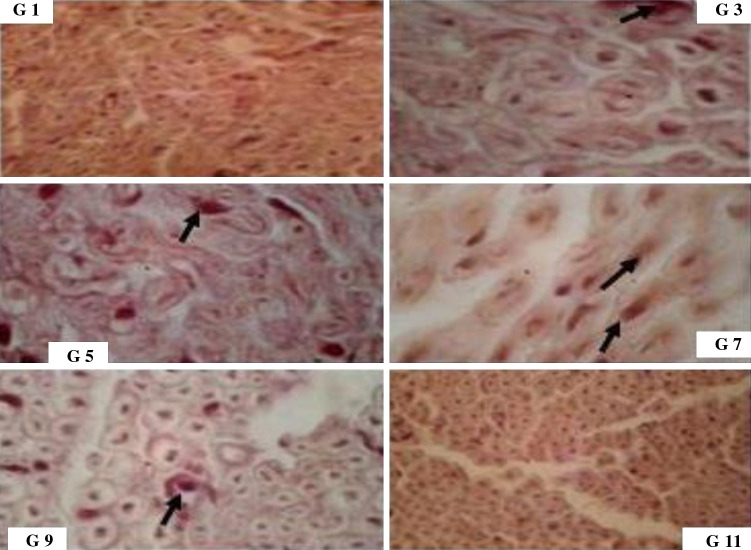
The present work was designed to investigate the potential protective effects of post-ischemic treatment with aminoguanidine (AG) on sciatic nerve ischemia/reperfusion (I/R) injury in rat. Seventy-two rats were divided into 12 groups (n = 6). We used ischemia model in these groups by occluding the right common iliac and femoral arteries for 3 h with a silk suture 6-0 using slipknot technique. Treatment groups (2, 4, 6, 8, 10, and 12) received 150 mg/kg AG intraperitoneally 24 h after induction of ischemia. After certain time intervals of reperfusion (2, 4, 7, 14, and 28 days), the function of the hind limb was assessed using behavioral scores based on gait, racing reflex, toe spread, pinch sensitivity, paw position, and grasp. After euthanasia, sciatic nerves were removed at the end of reperfusion times and sections were cut at 5 μm, then were stained for light microscopy studies and graded for ischemic fiber degeneration (IFD), edema, and apoptosis. Maximal behavioral deficit occurred at 7 days of reperfusion. The comparison of behavioral score pertaining to the control and AG groups revealed significant differences and showed also a better time course in recovery (P < 0.05). Other than 3 and 4 groups, the amount of edema in AG treatment groups showed significant differences compared with control groups (P < 0.05). IFD was also significantly decreased in the AG treatment groups than controls. Most importantly, I/R-induced apoptosis were improved significantly on the 4th, 7(th), and 14th days of reperfusion in AG-treated groups compared to controls. In conclusion, our findings suggest that post-ischemic administration of AG exhibits protective effect against sciatic nerve I/R injury.


Figures

Fig. 1
Fig. 1
Result of Kurskal–Wallis and Mann–Whitney U tests for behavioral score in different groups. Behavioral score (y-axis) for 3 h ischemia followed by certain time intervals of reperfusion (x-axis). There were significant differences between the I/R groups and normal group. Functions acquired were greater for the aminoguanidine groups than nontreated groups. a Control groups (aminoguanidine nontreated groups); b aminoguanidine-treated groups. Data are expressed as the median and interquartile range. **P < 0.01, for I/R groups vs. normal group. + P < 0.05, for I/R aminoguanidine treatment groups vs. I/R groups
Fig. 2
Fig. 2
Effects of aminoguanidine on edema grade for 3 h ischemia followed by certain time intervals of reperfusion. Data were analyzed by Kruskal–Wallis nonparametric one-way analysis of variance (ANOVA), followed by the Mann–Whitney U test for comparison edema in different groups. Except for the group 4 (2 day reperfusion), edema diminished significantly (P < 0.01) in AG-treated groups on days 4, 7, 14, and 28 reperfusion (a) compared with nontreatment groups (b). There also were significant differences between the I/R groups and normal group (P < 0.01). **P < 0.01, for I/R groups vs. normal group. ++ P < 0.01, for I/R aminoguanidine treatment groups vs. I/R groups
Fig. 3
Fig. 3
Effects of aminoguanidine on fiber degeneration (IFD) grade for 3 h ischemia followed by certain time intervals of reperfusion. Data were analyzed by Kruskal–Wallis nonparametric one-way analysis of variance (ANOVA), followed by the Mann–Whitney U test for comparison IFD in different groups. A significant increase in IFD was seen on days 2, 4, and 7 reperfusion (a). Maximum IFD were occurred on day 7 reperfusion. IFD decreased significantly in AG-treated groups on days 4 and 7 reperfusion (b) (P < 0.01). However, despite the expected, a significant increase in IFD was observed on day 2 reperfusion affected by aminoguanidine. *P < 0.01, for I/R groups vs. normal group. + P < 0.01, for I/R aminoguanidine treatment groups vs. I/R groups
Fig. 4
Fig. 4
Histopathological assessment of a transverse section of the sciatic nerve after 3 h ischemia followed by different time intervals of reperfusion (4, 7, and 14 days).The I/R groups (G5, G7, and G9) showed epineural edema and demyelination (Gomori trichrome stain). Epineural edema and demyelination decreased in the aminoguanidine-treated groups (G6, G8, and G10) (Gomori trichrome stain). Control: normal sciatic nerve, G group, dr days reperfusion
Fig. 5
Fig. 5
Comparison of apoptotic cells ratio (y-axis) in different groups (x-axis), for 3 h ischemia followed by certain time intervals of reperfusion (from 2 to 28 days). Values are mean ± SD (n = 6). Rate of apoptosis was significantly higher in all reperfusion periods, but maximum apoptosis were occurred in group 7 on day 7 reperfusion. Aminoguanidine significantly diminished apoptotic cells ratio in group 6 (4 day reperfusion) and group 8 (7 day reperfusion), *P < 0.05, for I/R groups vs. normal group. # P < 0.05, for I/R aminoguanidine treatment groups vs. I/R groups
Fig. 6
Fig. 6
Effects of 3 h ischemia followed by certain time intervals of reperfusion (from 2 to 28 days) on sciatic nerve apoptosis. Apoptosis was evaluated via TUNEL assay with the use of In Situ Cell Death Detection Kit at 40× magnification (arrows show the apoptotic cells)
Fig. 7
Fig. 7
Effects of aminoguanidine on sciatic nerve apoptosis for 3 h ischemia followed by certain time intervals of reperfusion (from 2 to 28 days). Apoptosis was evaluated via TUNEL assay with the use of In Situ Cell Death Detection Kit at 40× magnification (arrows show the apoptotic cells)

References

    1. Abdel-Rahman E, Bolton WK (2002) Pimagedine: a novel therapy for diabetic nephropathy. Expert Opin Investig Drugs 11:565–574 - PubMed
    1. Al-Majed AA, Neumann CM, Brushart TM, Gordon T (2000) Brief electrical stimulation promotes the speed and accuracy of motor axonal regeneration. J Neurosci 20:2602–2608 - PMC - PubMed
    1. Al-Majed AA, Khattab M, Raza M, Al-Shabanah OA, Mostafa AM (2003) Potentiation of diclofenac-induced anti-inflammatory response by aminoguanidine in carrageenan-induced acute inflammation in rats: the role of nitric oxide. Inflamm Res 52:378–382 - PubMed
    1. Anderson GM, Nukada H, McMorran PD (1997) Carbonyl histochemistry in rat reperfusion nerve injury. Brain Res 772:156–160 - PubMed
    1. Cash D, Beech JS, Rayne RC, Bath PM, Meldrum BS, Williams SC (2001) Neuroprotective effect of aminoguanidine on transient focal ischaemia in the rat brain. Brain Res 905:91–103 - PubMed

Publication types

MeSH terms

LinkOut - more resources