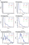
Three-dimensional radiobiological dosimetry (3D-RD) with 124I PET for 131I therapy of thyroid cancer
- PMID: 21484384
- PMCID: PMC3172686
- DOI: 10.1007/s00259-011-1769-1
Three-dimensional radiobiological dosimetry (3D-RD) with 124I PET for 131I therapy of thyroid cancer
Abstract
Radioiodine therapy of thyroid cancer was the first and remains among the most successful radiopharmaceutical (RPT) treatments of cancer although its clinical use is based on imprecise dosimetry. The positron emitting radioiodine, (124)I, in combination with positron emission tomography (PET)/CT has made it possible to measure the spatial distribution of radioiodine in tumors and normal organs at high resolution and sensitivity. The CT component of PET/CT has made it simpler to match the activity distribution to the corresponding anatomy. These developments have facilitated patient-specific dosimetry (PSD), utilizing software packages such as three-dimensional radiobiological dosimetry (3D-RD), which can account for individual patient differences in pharmacokinetics and anatomy. We highlight specific examples of such calculations and discuss the potential impact of (124)I PET/CT on thyroid cancer therapy.
Conflict of interest statement
Figures


Similar articles
-
Patient-specific dosimetry for 131I thyroid cancer therapy using 124I PET and 3-dimensional-internal dosimetry (3D-ID) software.J Nucl Med. 2004 Aug;45(8):1366-72. J Nucl Med. 2004. PMID: 15299063
-
Monte Carlo-based 3-dimensional dosimetry of salivary glands in radioiodine treatment of differentiated thyroid cancer estimated using 124I PET.Q J Nucl Med Mol Imaging. 2013 Mar;57(1):79-91. Q J Nucl Med Mol Imaging. 2013. PMID: 23474639 Free PMC article.
-
Acquisition settings for PET of 124I administered simultaneously with therapeutic amounts of 131I.J Nucl Med. 2006 Aug;47(8):1375-81. J Nucl Med. 2006. PMID: 16883019
-
Radioiodine therapy dosimetry in benign thyroid disease and differentiated thyroid carcinoma.Eur J Nucl Med Mol Imaging. 2010 Apr;37(4):821-8. doi: 10.1007/s00259-010-1398-0. Eur J Nucl Med Mol Imaging. 2010. PMID: 20186542 Review. No abstract available.
-
The role of (124)I-PET in diagnosis and treatment of thyroid carcinoma.Q J Nucl Med Mol Imaging. 2008 Mar;52(1):30-6. Q J Nucl Med Mol Imaging. 2008. PMID: 17657202 Review.
Cited by
-
VIDA: a voxel-based dosimetry method for targeted radionuclide therapy using Geant4.Cancer Biother Radiopharm. 2015 Feb;30(1):16-26. doi: 10.1089/cbr.2014.1713. Epub 2015 Jan 16. Cancer Biother Radiopharm. 2015. PMID: 25594357 Free PMC article.
-
I-124 Imaging and Dosimetry.Mol Imaging Radionucl Ther. 2017 Feb 9;26(Suppl 1):66-73. doi: 10.4274/2017.26.suppl.07. Mol Imaging Radionucl Ther. 2017. PMID: 28117290 Free PMC article.
-
Radionuclide therapy: current status and prospects for internal dosimetry in individualized therapeutic planning.Clinics (Sao Paulo). 2019;74:e835. doi: 10.6061/clinics/2019/e835. Epub 2019 Jul 29. Clinics (Sao Paulo). 2019. PMID: 31365617 Free PMC article. Review.
-
Thyroid Cancer Radiotheragnostics: the case for activity adjusted 131I therapy.Clin Transl Imaging. 2018 Oct;6(5):335-346. doi: 10.1007/s40336-018-0291-x. Epub 2018 Aug 4. Clin Transl Imaging. 2018. PMID: 30911535 Free PMC article.
-
Tumor Response to Radiopharmaceutical Therapies: The Knowns and the Unknowns.J Nucl Med. 2021 Dec;62(Suppl 3):12S-22S. doi: 10.2967/jnumed.121.262750. J Nucl Med. 2021. PMID: 34857617 Free PMC article. Review.
References
-
- Hellman S, Devita VT, Rosenberg SA. Cancer: principles & practice of oncology. 6. Philadelphia: Lippincott Williams and Wilkins; 2001. Principles of cancer management: radiation therapy; pp. 265–88.
-
- Cooper DS, Doherty GM, Haugen BR, Kloos RT, Lee SL, et al. American Thyroid Association (ATA) Guidelines Taskforce on Thyroid Nodules and Differentiated Thyroid Cancer. Revised American Thyroid Association management guidelines for patients with thyroid nodules and differentiated thyroid cancer. Thyroid. 2009;19(11):1167–214. - PubMed
-
- Van Nostrand D, Atkins F, Yeganeh F, Acio E, Bursaw R, Wartofsky L. Dosimetrically determined doses of radioiodine for the treatment of metastatic thyroid carcinoma. Thyroid. 2002;12 (2):121–34. - PubMed
-
- Holst JP, Burman KD, Atkins F, Umans JG, Jonklaas J. Radio-iodine therapy for thyroid cancer and hyperthyroidism in patients with end-stage renal disease on hemodialysis. Thyroid. 2005;15 (12):1321–31. - PubMed
-
- Driedger AA, Quirk S, McDonald TJ, Ledger S, Gray D, Wall W, et al. A pragmatic protocol for I-131 rhTSH-stimulated ablation therapy in patients with renal failure. Clin Nucl Med. 2006;31(8):454–7. - PubMed
Publication types
MeSH terms
Substances
Grants and funding
LinkOut - more resources
Full Text Sources
Other Literature Sources
Medical
Miscellaneous

