Quantitative imaging of 124I and 86Y with PET
- PMID: 21484385
- PMCID: PMC3098993
- DOI: 10.1007/s00259-011-1768-2
Quantitative imaging of 124I and 86Y with PET
Abstract
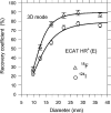
The quantitative accuracy and image quality of positron emission tomography (PET) measurements with (124)I and (86)Y is affected by the prompt emission of gamma radiation and positrons in their decays, as well as the higher energy of the emitted positrons compared to those emitted by (18)F. PET scanners cannot distinguish between true coincidences, involving two 511-keV annihilation photons, and coincidences involving one annihilation photon and a prompt gamma, if the energy of this prompt gamma is within the energy window of the scanner. The current review deals with a number of aspects of the challenge this poses for quantitative PET imaging. First, the effect of prompt gamma coincidences on quantitative accuracy of PET images is discussed and a number of suggested corrections are described. Then, the effect of prompt gamma coincidences and the increased singles count rates due to gamma radiation on the count rate performance of PET is addressed, as well as possible improvements based on modification of the scanner's energy windows. Finally, the effect of positron energy on spatial resolution and recovery is assessed. The methods presented in this overview aim to overcome the challenges associated with the decay characteristics of (124)I and (86)Y. Careful application of the presented correction methods can allow for quantitatively accurate images with improved image contrast.
Figures


Similar articles
-
Three-dimensional positron emission tomography imaging with 124I and 86Y.Nucl Med Commun. 2006 Mar;27(3):237-45. doi: 10.1097/01.mnm.0000199476.46525.2c. Nucl Med Commun. 2006. PMID: 16479243
-
Quantitative imaging and correction for cascade gamma radiation of 76Br with 2D and 3D PET.Phys Med Biol. 2002 Oct 7;47(19):3519-34. doi: 10.1088/0031-9155/47/19/306. Phys Med Biol. 2002. PMID: 12408479
-
Simulation of triple coincidences in PET.Phys Med Biol. 2015 Jan 7;60(1):117-36. doi: 10.1088/0031-9155/60/1/117. Epub 2014 Dec 5. Phys Med Biol. 2015. PMID: 25479147
-
PET imaging problems with the non-standard positron emitters Yttrium-86 and Iodine-124.Q J Nucl Med Mol Imaging. 2008 Jun;52(2):159-65. Epub 2007 Nov 28. Q J Nucl Med Mol Imaging. 2008. PMID: 18043538 Review.
-
X-ray-based attenuation correction for positron emission tomography/computed tomography scanners.Semin Nucl Med. 2003 Jul;33(3):166-79. doi: 10.1053/snuc.2003.127307. Semin Nucl Med. 2003. PMID: 12931319 Review.
Cited by
-
Multimodal Imaging Technology Effectively Monitors HER2 Expression in Tumors Using Trastuzumab-Coupled Organic Nanoparticles in Patient-Derived Xenograft Mice Models.Front Oncol. 2021 Nov 17;11:778728. doi: 10.3389/fonc.2021.778728. eCollection 2021. Front Oncol. 2021. PMID: 34869025 Free PMC article.
-
Cerenkov luminescence and PET imaging of 90Y: capabilities and limitations in small animal applications.Phys Med Biol. 2020 Mar 20;65(6):065006. doi: 10.1088/1361-6560/ab7502. Phys Med Biol. 2020. PMID: 32045899 Free PMC article.
-
Simultaneous quantitative imaging of two PET radiotracers via the detection of positron-electron annihilation and prompt gamma emissions.Nat Biomed Eng. 2023 Aug;7(8):1028-1039. doi: 10.1038/s41551-023-01060-y. Epub 2023 Jul 3. Nat Biomed Eng. 2023. PMID: 37400715 Free PMC article.
-
Quantification, improvement, and harmonization of small lesion detection with state-of-the-art PET.Eur J Nucl Med Mol Imaging. 2017 Aug;44(Suppl 1):4-16. doi: 10.1007/s00259-017-3727-z. Epub 2017 Jul 8. Eur J Nucl Med Mol Imaging. 2017. PMID: 28687866 Free PMC article. Review.
-
Efficiency of 124I radioisotope production from natural and enriched tellurium dioxide using 124Te(p,xn)124I reaction.EJNMMI Phys. 2022 Jun 6;9(1):41. doi: 10.1186/s40658-022-00471-1. EJNMMI Phys. 2022. PMID: 35666325 Free PMC article.
References
-
- Perk LR, Visser GW, Vosjan MJ, Stigter-van Walsum M, Tijink BM, Leemans CR, et al. (89)Zr as a PET surrogate radioisotope for scouting biodistribution of the therapeutic radiometals (90)Y and (177)Lu in tumor-bearing nude mice after coupling to the internalizing antibody cetuximab. J Nucl Med. 2005;46(11):1898–1906. - PubMed
Publication types
MeSH terms
Substances
LinkOut - more resources
Full Text Sources
Other Literature Sources

