. 2011 May;19(4):457-70.
doi: 10.1007/s10577-011-9208-5. Epub 2011 Apr 12.

Genomic size of CENP-A domain is proportional to total alpha satellite array size at human centromeres and expands in cancer cells

Affiliations

Genomic size of CENP-A domain is proportional to total alpha satellite array size at human centromeres and expands in cancer cells

Lori L Sullivan et al. Chromosome Res. 2011 May.

Abstract

Human centromeres contain multi-megabase-sized arrays of alpha satellite DNA, a family of satellite DNA repeats based on a tandemly arranged 171 bp monomer. The centromere-specific histone protein CENP-A is assembled on alpha satellite DNA within the primary constriction, but does not extend along its entire length. CENP-A domains have been estimated to extend over 2,500 kb of alpha satellite DNA. However, these estimates do not take into account inter-individual variation in alpha satellite array sizes on homologous chromosomes and among different chromosomes. We defined the genomic distance of CENP-A chromatin on human chromosomes X and Y from different individuals. CENP-A chromatin occupied different genomic intervals on different chromosomes, but despite inter-chromosomal and inter-individual array size variation, the ratio of CENP-A to total alpha satellite DNA size remained consistent. Changes in the ratio of alpha satellite array size to CENP-A domain size were observed when CENP-A was overexpressed and when primary cells were transformed by disrupting interactions between the tumor suppressor protein Rb and chromatin. Our data support a model for centromeric domain organization in which the genomic limits of CENP-A chromatin varies on different human chromosomes, and imply that alpha satellite array size may be a more prominent predictor of CENP-A incorporation than chromosome size. In addition, our results also suggest that cancer transformation and amounts of centromeric heterochromatin have notable effects on the amount of alpha satellite that is associated with CENP-A chromatin.


Figures
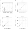
Figure 1
Figure 1. Range of centromeric fiber stretching and relationship between CENP-A immunofluorescence and centromeric fiber length
(a) Comparison of chromatin fiber resolution (kb/μm) in a representative euchromatic region (HSA6) versus a centromere (DXZ1) showed that there was more variability in stretching within centromeric regions. When only long centromeric fibers (i.e. those that spanned multiple fields of view) were exclusively analyzed, euchromatic and centromeric regions were stretched to similar extents. Graphs show the linear relationship between increasing fiber length and fluorescence for centromeres. CENP-A immunofluorescence (y-axis) and alpha satellite fluorescence (x-axis) was plotted for DXZ1 in two representative cell lines (b) DIP1 and (c) DIP3 and for (d) DYZ3 in a third cell line DIP4. Linear regression was performed using Kaleidagraph software. The R2 values and equation for the line are indicated on each graph.
Figure 2
Figure 2. Alpha satellite array size and CENP-A domain size at centromeres of chromosomes X and Y
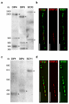
Molecular sizes of alpha satellite arrays were determined by pulsed field gel electrophoresis (PFGE) of high molecular weight DNA embedded in agarose, followed by non-radioactive Southern blotting. (a) Sizes of X alpha satellite (DXZ1) arrays in two diploid lines (DIP4, DIP5) and a mouse-human somatic cell hybrid containing a single human X chromosome (SCX3) ranged from 3.7-4.2Mb. The total array size for DXZ1 in each cell line was estimated by adding the molecular weights of all bands that appeared in the Southern blot. Digestion of DNA embedded within agarose plugs with BglI (shown) or BstEII (not shown) released DXZ1 as two or three bands that were detected by Southern blotting. High molecular weight bands are denoted with asterisks (*). PFGE conditions for DXZ1 were 3 V/cm, initial switch time of 250s, and a final switch time of 900s, for 50 hours. The figure shows three cell lines that were run on separate but identically sized gels under identical conditions using the same high molecular weight standards. The gel for each cell line was cropped identically and aligned according to the molecular weight standards. (b) CENP-A domains sizes were determined using CENP-A immunostaining on extended chromatin fibers followed by FISH with a DNA probe specific for DXZ1. Domain sizes were calculated by assigning molecular sizes as determined by PFGE to the length of the alpha satellite signals (in micrometers). CENP-A domains were calculated by comparing the length of the immunostaining on the fiber to the length of the alpha satellite fluorescent signal (see Materials and Methods for additional details). The representative image from cell line DIP3 shows CENP-A (red) immunostaining overlapping with only a portion of DXZ1 (green). The merged image is shown on the left, followed by individual image channels for CENP-A (middle) and DXZ1 (right). Scale bar is 10 micrometers. (c) Molecular sizing of Y alpha satellite (DYZ3) arrays in two diploid lines (DIP1, DIP4) and a mouse-human somatic cell hybrid containing a single human Y chromosome (SCY1) showed arrays that ranged from 350-600kb. Digestion of DNA embedded within agarose plugs with BamHI released the entire DYZ3 array that was detected by Southern blotting as a single band. High molecular weight bands are denoted with asterisks (*). PFGE parameters were 6 V/cm, 10s initial switch time, 80s final switch time, for 23 hours. The figure shows three cell lines that were run on separate but identically sized gels under identical conditions and using the same high molecular weight standards. The gel for each cell line was cropped identically and aligned according to the molecular weight standards. (d) Representative image from cell line DIP4 showing CENP-A (red) immunostaining overlapping with a portion of DYZ3 (green). The merged image is shown on the left, followed by individual image channels for CENP-A (middle) and DYZ3 (right). Scale bar is 5 micrometers.
Figure 3
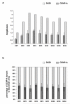
Figure 3. Human chromosome X (HSAX) alpha satellite (DXZ1) array size compared to CENP-A chromatin domain size
(a) DXZ1 arrays (light gray bars) were measured on different HSAXs present in human diploid (DIP) male lines or rodent-human somatic cell hybrids (SCX) in which HSAX was the only human chromosome. CENP-A chromatin domain size in megabases (dark gray bars) was determined using CENP-A immunostaining and FISH with a DXZ1 probe. DXZ1 arrays and CENP-A domain sizes varied up to three-fold among individual HSAXs. Error bars represent standard deviations in the genomic length (in megabases). (b) Calculation of the percentage of DXZ1 occupied by the CENP-A domain (dark gray bars) showed that the CENP-A domain was proportionally assembled on DXZ1 arrays (light gray bars) that varied in size among different HSAXs. Thirty-five to forty-five percent of DXZ1 was occupied by CENP-A chromatin. Error bars represent standard deviations in the percentage of DXZ1 occupied by the CENP-A domain.
Figure 4
Figure 4. Human chromosome Y (HSAY) alpha satellite (DYZ3) array size compared to CENP-A chromatin domain size
(a) DYZ3 arrays (light gray bars) were measured on different HSAYs present in human diploid (DIP) male lines or rodent-human somatic cell hybrids (SCY) in which HSAY was the only human chromosome. CENP-A chromatin domain size in megabases (dark gray bars) was determined using CENP-A immunostaining and FISH with a DYZ3 probe. As previously reported for DYZ3, arrays were distributed into two size groups: those <600kb and those >1Mb (Abruzzo et al., 1996). Accordingly, CENP-A domains clustered into groups that were either <200kb or >500kb. Error bars represent standard deviations in the genomic length (in megabases). (b) Calculation of the percentage of DYZ3 occupied by the CENP-A domain (dark gray bars) showed that the CENP-A domain was proportionally assembled on DYZ3 (light gray bars) that varied in size among different HSAYs. Typically, 45-50% of DYZ3 was occupied by CENP-A chromatin. Error bars represent standard deviations in the percentage of DYZ3 occupied by the CENP-A domain.
Figure 5
Figure 5. CENP-A domains size changes in response to protein dosage
(a) Primary cell line (DIP3) was transformed by virally-expressing HPV E7 oncoprotein (DIP3-E7). E7 binds to retinoblastoma protein (Rb) and inactivates it, preventing it from binding to its nuclear targets. Western blotting with anti-Rb antibodies demonstrated that the amount of chromatin-bound Rb was undetectable upon E7 expression and transformation of DIP3. (b) ChIP-PCR showed that in the absence of chromatin-bound Rb, heterochromatic histone modifications H3K9me2, H3K9me3 and H4K20me3 at DXZ1 were decreased. The poised euchromatic mark H3K4me2 was not significantly different at DXZ1 in DIP3 before and after transformation. Enrichment at DXZ1 was calculated as the ratio of immunoprecipitated DNA to input. GAPDH and AFM were used as genic control sites. Error bars represent standard deviations. (c) CENP-A chromatin domain size at DXZ1 was measured using CENP-A immunostaining and FISH in cells over-expressing CENP-A (DIP2-OE) and after HPV-E7-mediated transformation/heterochromatin depletion of a primary fibroblast line DIP3 to create DIP3-E7. Comparisons in CENP-A domain size were made to the parent line (i.e. DIP2 vs DIP2-OE and DIP3 vs DIP3-E7). In each case, CENP-A domain size on DXZ1 increased by 1.5 fold of its original size. Error bars represent standard deviations in the total number of megabases occupied by the CENP-A chromatin domain. (d) CENP-A chromatin domain size on DYZ3 was measured as in (c) in cell lines DIP2-OE and DIP3-E7. Comparisons were made to the parental line in each case (i.e. DIP2 vs DIP2-OE and DIP3 vs DIP3-E7). The domain increased 1.5 times in length over DYZ3 when CENP-A was over-expressed or heterochromatin was depleted from the centromere. Error bars represent standard deviations in the total number of megabases occupied by the CENP-A chromatin domain. Significant differences in (b), (c) and (d) were calculated using a Student’s t-test.

Similar articles

Cited by

References

    1. ABRUZZO MA, GRIFFIN DK, MILLIE EA, SHEEAN LA, HASSOLD TJ. The effect of Y-chromosome alpha-satellite array length on the rate of sex chromosome disomy in human sperm. Hum Genet. 1996;97:819–23. - PubMed
    1. ALONSO A, FRITZ B, HASSON D, ABRUSAN G, CHEUNG F, YODA K, RADLWIMMER B, LADURNER AG, WARBURTON PE. Co-localization of CENP-C and CENP-H to discontinuous domains of CENP-A chromatin at human neocentromeres. Genome Biol. 2007;8:R148. - PMC - PubMed
    1. ALONSO A, HASSON D, CHEUNG F, WARBURTON PE. A paucity of heterochromatin at functional human neocentromeres. Epigenetics Chromatin. 2010;3:6. - PMC - PubMed
    1. ALONSO A, MAHMOOD R, LI S, CHEUNG F, YODA K, WARBURTON PE. Genomic microarray analysis reveals distinct locations for the CENP-A binding domains in three human chromosome 13q32 neocentromeres. Hum Mol Genet. 2003;12:2711–21. - PubMed
    1. AMATO A, SCHILLACI T, LENTINI L, DI LEONARDO A. CENPA overexpression promotes genome instability in pRb-depleted human cells. Mol Cancer. 2009;8:119. - PMC - PubMed

Publication types

LinkOut - more resources