Review
. 2011 Sep;469(9):2440-51.
doi: 10.1007/s11999-011-1892-0.

How reverse shoulder arthroplasty works

Affiliations
Review

How reverse shoulder arthroplasty works

Matthew Walker et al. Clin Orthop Relat Res. 2011 Sep.

Abstract

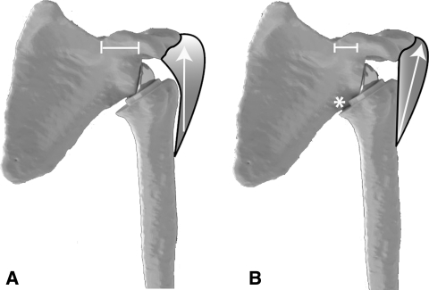
Background: The reverse total shoulder arthroplasty was introduced to treat the rotator cuff-deficient shoulder. Since its introduction, an improved understanding of the biomechanics of rotator cuff deficiency and reverse shoulder arthroplasty has facilitated the development of modern reverse arthroplasty designs.

Questions/purposes: We review (1) the basic biomechanical challenges associated with the rotator cuff-deficient shoulder; (2) the biomechanical rationale for newer reverse shoulder arthroplasty designs; (3) the current scientific evidence related to the function and performance of reverse shoulder arthroplasty; and (4) specific technical aspects of reverse shoulder arthroplasty.

Methods: A PubMed search of the English language literature was conducted using the key words reverse shoulder arthroplasty, rotator cuff arthropathy, and biomechanics of reverse shoulder arthroplasty. Articles were excluded if the content fell outside of the biomechanics of these topics, leaving the 66 articles included in this review.

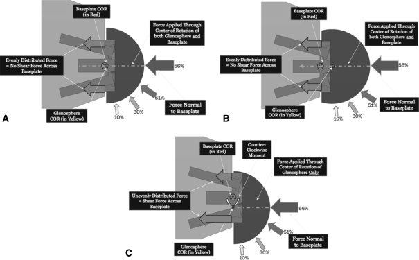
Results: Various implant design factors as well as various surgical implantation techniques affect stability of reverse shoulder arthroplasty and patient function. To understand the implications of individual design factors, one must understand the function of the normal and the cuff-deficient shoulder and coalesce this understanding with the pathology presented by each patient to choose the proper surgical technique for reconstruction.

Conclusions: Several basic science and clinical studies improve our understanding of various design factors in reverse shoulder arthroplasty. However, much work remains to further elucidate the performance of newer designs and to evaluate patient outcomes using validated instruments such as the American Society for Elbow Surgery, simple shoulder test, and the Constant-Murley scores.


Figures

Fig. 1
Fig. 1
True AP (Grashey view [46]) of a shoulder with cuff tear arthropathy is shown. The film demonstrates glenohumeral arthritis, superior glenoid wear, proximal humeral migration, and acetabularization of the acromion.
Fig. 2
Fig. 2
This figure demonstrates the wide spectrum of disease that can be seen in rotator cuff tear arthropathy.
Fig. 3
Fig. 3
This figure shows the Delta 3 reverse arthroplasty.
Fig. 4
Fig. 4
This artist rendition uses billiard balls to explain concavity-compression. Deeper concavities require a larger displacing force for a given compressive load. Reprinted with permission from Chapter 8, Principles of glenohumeral stability. In: Matsen F, Lippitt S, eds. Shoulder Surgery: Principles and Procedures. Philadelphia: WB Saunders; 2003, Fig. 8-5.
Fig. 5A–D
Fig. 5A–D
(A) This computer model demonstrates the anterior view of the glenoid center line. (B) This computer model demonstrates the inferior view of the glenoid center line. (C) This computer model demonstrates the anterior view of the alternate center line along the scapular spine. (D) This computer model demonstrates the inferior view of the alternate center line along the scapular spine.
Fig. 6
Fig. 6
The DJO RSP has a central 6.5-mm lag screw.
Fig. 7A–C
Fig. 7A–C
(A) The figure shows the progression of joint forces throughout abduction for the concentric glenosphere. (B) This figure shows the progression of joint forces throughout abduction for the lateral eccentric glenosphere. (C) This figure shows the progression of joint forces throughout abduction for the inferior eccentric glenosphere. Reprinted with permission from Gutiérrez S, Walker M, Willis M, Pupello D, Frankle M. Effects of tilt and glenosphere eccentricity on baseplate/bone interface forces in a computational model, validated by a mechanical model, of reverse shoulder arthroplasty. Figure 5. J Shoulder Elbow Surg. 2011 Jan 31 [Epub ahead of print].
Fig. 8
Fig. 8
This figure shows the effect of different forces at the baseplate-bone interface.
Fig. 9A–B
Fig. 9A–B
(A) Devices with lateralized centers of rotation allow the proximal pull of the deltoid to pull the humerus toward the glenosphere, thus increasing the compression and stability of the implant. (B) Devices with medialized centers of rotation allow the deltoid to place a distraction moment on the prosthesis and increase the propensity for dislocation.
Fig. 10
Fig. 10
The balance stability angle. This represents the maximum angle that the net force on the humeral head forms with the glenoid centerline before dislocation. Reprinted with permission from Chapter 9, Principles of the glenoid concavity. In: Matsen F, Lippitt S, eds. Shoulder Surgery: Principles and Procedures. Philadelphia: WB Saunders; 2003, Fig. 9-23.

References

    1. Anglin C, Wyss UP, Pichore DR. Shoulder prosthesis subluxation: theory and experiment. J Shoulder Elbow Surg. 2000;9:104–114. doi: 10.1016/S1058-2746(00)90038-7. - DOI - PubMed
    1. Bayley J, Kessel L. The Kessel total shoulder replacement. In: Bayley I, Kessel L, editors. Shoulder Surgery. New York: Springer-Verlag; 1982. pp. 160–164.
    1. Bobyn JD, Toh K-K, Hacking SA, Tanzer M, Krygier JJ. Tissue response to porous tantalum acetabular cups: a canine model. J Arthroplasty. 1999;14:347–354. doi: 10.1016/S0883-5403(99)90062-1. - DOI - PubMed
    1. Boileau P, Chuinard C, Roussanne Y, Bicknell RT, Rochet N, Trojani C. Reverse shoulder arthroplasty combined with a modified latissimus dorsi and teres major tendon transfer for shoulder pseudoparalysis associated with dropping arm. Clin Orthop Relat Res. 2008;466:584–593. doi: 10.1007/s11999-008-0114-x. - DOI - PMC - PubMed
    1. Boileau P, Chuinard C, Roussanne Y, Neyton L, Trojani C. Modified latissimus dorsi and teres major transfer through a single delto-pectoral approach for external rotation deficit of the shoulder: as an isolated procedure or with a reverse arthroplasty. J Shoulder Elbow Surg. 2007;16:671–682. doi: 10.1016/j.jse.2007.02.127. - DOI - PubMed

MeSH terms