. 2011 Jul;41(7):1862-71.
doi: 10.1002/eji.201041195. Epub 2011 Jun 7.

TSLP enhances the function of helper type 2 cells

Affiliations

TSLP enhances the function of helper type 2 cells

Masayuki Kitajima et al. Eur J Immunol. 2011 Jul.

Abstract

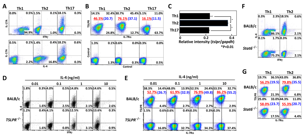
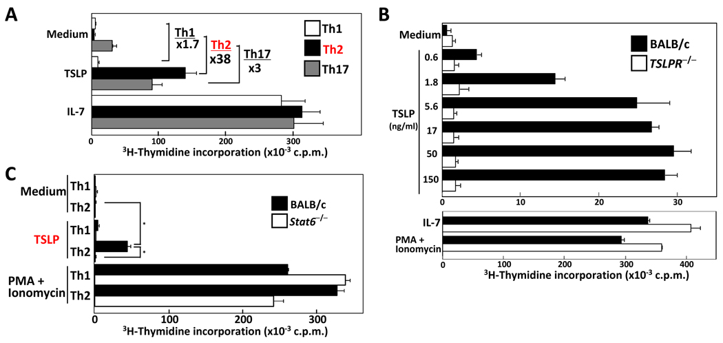
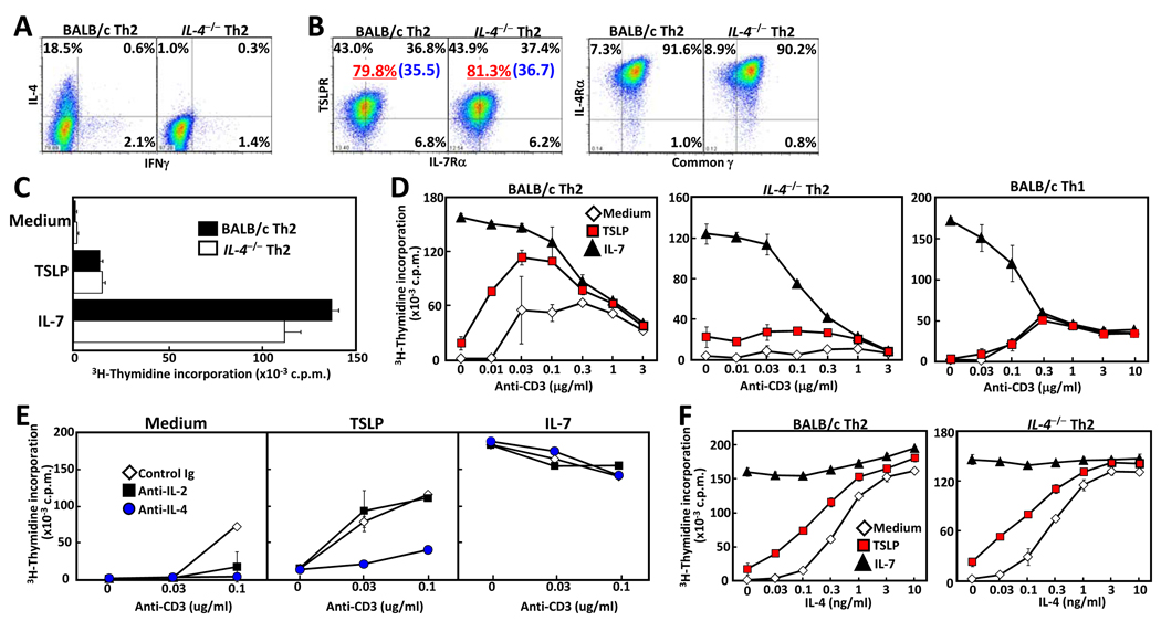
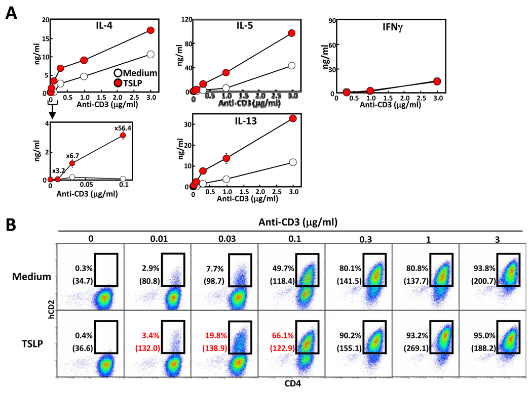
The cytokine thymic stromal lymphopoietin (TSLP) has been implicated in the development and progression of allergic inflammation in both humans and mice. TSLP has been shown to promote a Th2-type response through upregulation of OX40L on dendritic cells, and through direct induction of IL-4 production in naïve CD4+ T cells. However, its direct effect on effector Th cells has not been extensively investigated. In this study, we show that the level of TSLP receptor (TSLPR) expression on mouse effector Th2 cells is higher than on Th1 and Th17 cells, and that TSLP induced proliferation of effector Th2, but not Th1 nor Th17 cells. TSLP also induced the phosphorylation of signal transducer and activator of transcription (Stat) 5, and expression of the anti-apoptotic factor Bcl-2 in Th2 cells. Finally, TSLP-mediated proliferation on Th2 cells was enhanced by TCR stimulation, through IL-4-mediated induction of TSLPR expression. Taken together, these results indicate that TSLP is involved in exacerbation of mouse Th2-mediated allergic inflammation in a Th2 environment through direct stimulation of Th2 effector cells.


Conflict of interest statement

Conflict of interest

The authors declare no financial or commercial conflict of interest.

Figures

Figure 1
Figure 1. Increased TSLPR expression on Th2 cells by IL-4-Stat6 dependent pathway
CD4+CD62hi T cells from BALB/c mice were stimulated with immobilized anti-CD3 mAb and anti-CD28 mAb under Th1/Th2/Th17 conditions for 5 days. (A) Cells were re-stimulated by anti-CD3 mAbs for 6hr and then assessed for IFNγ, IL-4 and IL-17 production by intracellular staining. (B) Cell-surface levels of TSLPR and IL-7Rα were measured by flow cytometry and compared to isotype staining samples. (C) mRNA levels of tslpr and gapdh were determined by quantitative RT-PCR. The relative intensity (/gapdh) is shown with standard deviation ( n=3). (D, E) CD4+CD62hi T cells from BALB/c and TSLPR−/−mice were differentiated into Th2 cells and cultured in varying doses of IL-4 for 5 days. (F, G) CD4+CD62hi T cells from BALB/c and Stat6−/−mice were differentiated into Th1 orTh2 cells. Cells were measured for IFNγ and IL-4 production by intracellular staining (D, F), and for cell-surface expression of TSLPR and IL-7Rα (E, G). The underlined value represents the percentage of TSLPR+ cells and the value in parenthesis represents the Mean Fluorescence Intensity of TSLPR+ cells. Three independent experiments were performed with similar results. *p<0.01 by Student’s t test.
Figure 2
Figure 2. TSLP induces proliferation of Th2 cells
(A) Naïve CD62LhiCD4 T cells were differentiated into Th1, Th2, and Th17 lineages. The cells were cultured with medium, TSLP (50ng/ml), or IL-7 (10ng/ml) for 24 hr, then pulsed with 3H-thymidine for an additional 16 h and proliferation measured. (B) Wild-type and TSLPR−/−Th2 cells and (C) BALB/c and Stat6−/−Th1/Th2 cells were cultured in the presence or absence of the indicated concentration of TSLP, IL-7, or with PMA +ionomycin, and proliferation measured as in panel A. Data shown are mean+/− SD of threeindependent experiments. *p<0.01 by Student’s t test.
Figure 3
Figure 3. IL-4 enhances TSLP-mediated co-stimulation of Th2 cells
Naïve CD62LhiCD4 T cells from BALB/c and IL-4−/−mice were differentiated under Th2 conditions for 5 days. After washing of cells, cytokine production was assessed by intracellular staining (A). (B) TSLPR, IL-7Rα, IL-4Rα, and common γ chain expression were detected by flow cytometry. The underlined value represents the percentage of TSLPR+ cells and the value in parenthesis represents the Mean Fluorescence Intensity of TSLPR+ cells. (C) Proliferation was measured by 3H-thymidine incorporation following culture with medium, TSLP, or IL-7. (D) Wild-type and IL-4−/−Th2, and wild-type Th1 cells were cultured with medium, TSLP, or IL-7 in the presence or absence of plate-bound anti-CD3 (at indicated concentration) and proliferation was measured by 3H-thymidine incorporation. (E) Wild-type Th2 cells were cultured with medium, TSLP, or IL-7 with either control Ig, anti-IL-2, or anti-IL-4 Abs (each 2mg / well), and proliferation was measured by 3H-thymidine incorporation. (F) Wild-type and IL-4−/−Th2 cells were cultured with medium, TSLP, or IL-7, in the presence or absence of increasing amounts of IL-4, and proliferation was measured by 3H-thymidine incorporation. Data shown are mean +/− SD of three independent experiments.
Figure 4
Figure 4. Enhanced IL-4 production by TSLP co-stimulation
Effector Th2 cells were cultured with medium or TSLP in the presence or absence of increasing plate-bound anti-CD3 antibody. (A) After 48 hours of culture IL-4, IL-5, IL-13 and IFNγ concentration in the culture supernatant was measured by ELISA. (B) KN2/KN2 Th2 cells were cultured with medium or TSLP in the presence or absence of increasing anti-CD3 antibody for 24 hours, then stained with human CD2 and mouse CD4 antibodies and analyzed by flow cytometry. The value in parenthesis represents the Mean Fluorescence Intensity of CD4+ KN2+ cells. Data shown are representative of three independent experiments.
Figure 5
Figure 5. Enhanced TSLPR expression by IL-4 treatment on Th1, Th2, and Th17 cells
(A) Wild-type and IL-4−/− Th2 cells were cultured in medium alone or with TSLP in the presence or absence of plate-bound anti-CD3 (at indicated concentration) for 24 hours. (B) Wild-type Th2 cells were cultured in medium alone or with TSLP in the presence or absence of IL-4 (at indicated concentration) for 24 hours. (C) Th1, Th2, and Th17 cells were cultured in medium alone or with TSLP in the presence or absence of IL-4 for 24 hours. Then, TSLPR and IL-7Rα expression was detected by flow cytometry. The underlined value represents the percentage of TSLPR+ cells and the value in parenthesis represents the Mean Fluorescence Intensity of TSLPR+ cells. Data shown are representative of three independent experiments.

References

    1. Ziegler SF, Artis D. Sensing the outside world: TSLP regulates barrier immunity. Nature Immunology. 2010;11:289–293. - PMC - PubMed
    1. Soumelis V, Reche PA, Kanzler H, Yuan W, Edward G, Homey B, Gilliet M, Ho S, Antonenko S, Lauerma A, Smith K, Gorman D, Zurawski S, Abrams J, Menon S, McClanahan T, de Waal-Malefyt Rd R, Bazan F, Kastelein RA, Liu YJ. Human epithelial cells trigger dendritic cell mediated allergic inflammation by producing TSLP. Nature immunology. 2002;3:673–680. - PubMed
    1. Ying S, O'Connor B, Ratoff J, Meng Q, Mallett K, Cousins D, Robinson D, Zhang G, Zhao J, Lee TH, Corrigan C. Thymic stromal lymphopoietin expression is increased in asthmatic airways and correlates with expression of Th2-attracting chemokines and disease severity. The Journal of Immunology. 2005;174:8183–8190. - PubMed
    1. Headley MB, Zhou B, Shih WX, Aye T, Comeau MR, Ziegler SF. TSLP conditions the lung immune environment for the generation of pathogenic innate and antigen-specific adaptive immune responses. The Journal of Immunology. 2009;182:1641–1647. - PMC - PubMed
    1. Jessup HK, Brewer AW, Omori M, Rickel EA, Budelsky AL, Yoon BR, Ziegler SF, Comeau MR. Intradermal administration of thymic stromal lymphopoietin induces a T cell- and eosinophil-dependent systemic Th2 inflammatory response. The Journal of Immunology. 2008;181:4311–4319. - PubMed

Publication types

MeSH terms

LinkOut - more resources