

Thalamic-insular dysconnectivity in schizophrenia: evidence from structural equation modeling
- PMID: 21484952
- PMCID: PMC6870155
- DOI: 10.1002/hbm.21246
Thalamic-insular dysconnectivity in schizophrenia: evidence from structural equation modeling
Abstract
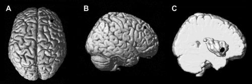
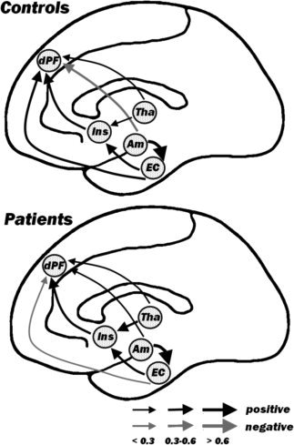
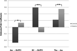
Structural and functional studies have shown that schizophrenia is often associated with frontolimbic abnormalities in the prefrontal and mediotemporal regions. It is still unclear, however, if such dysfunctional interaction extends as well to relay regions such as the thalamus and the anterior insula. Here, we measured gray matter volumes of five right-hemisphere regions in 68 patients with schizophrenia and 77 matched healthy subjects. The regions were amygdala, thalamus, and entorhinal cortex (identified as anomalous by prior studies on the same population) and dorsolateral prefrontal cortex and anterior insula (isolated by voxel-based morphometry analysis). We used structural equation modeling and found altered path coefficients connecting the thalamus to the anterior insula, the amygdala to the DLPFC, and the entorhinal cortex to the DLPFC. In particular, patients exhibited a stronger thalamus-insular connection than healthy controls. Instead, controls showed positive entorhinal-DLPFC and negative amygdalar-DLPFC connections, both of which were absent in the clinical population. Our data provide evidence that schizophrenia is characterized by an impaired right-hemisphere network, in which intrahemispheric communication involving relay structures may play a major role in sustaining the pathophysiology of the disease.
Copyright © 2011 Wiley Periodicals, Inc.
Figures
References
-
- Agarwal N, Rambaldelli G, Perlini C, Dusi N, Kitis O, Bellani M, Cerini R, Isola M, Versace A, Balestrieri M, Gasparini A, Mucelli RP, Tansella M, Brambilla P ( 2008): Microstructural thalamic changes in schizophrenia: A combined anatomic and diffusion weighted magnetic resonance imaging study. J Psychiatry Neurosci 33: 440–448. - PMC - PubMed
-
- Allen GV, Saper CB, Hurley KM, Cechetto DF ( 1991): Organization of visceral and limbic connections in the insular cortex of the rat. J Comp Neurol 311: 1–16. - PubMed
-
- American Psychiatric Association ( 2000): Diagnostic and Statistical Manual of Mental Disorders DSM‐IV‐TR, 4th ed Washington DC: American Psychiatric Publishing, Inc; Joreskog and Wold, 1982.
-
- Amunts K, Kedo O, Kindler M, Pieperhoff P, Mohlberg H, Shah NJ, Habel U, Schneider F, Zilles K ( 2005): Cytoarchitectonic mapping of the human amygdala, hippocampal region and entorhinal cortex: Intersubject variability and probability maps. Anat Embryol 210: 343–352. - PubMed
-
- Andreasen NC, Arndt S, Swayze V, Cizadlo T, Flaum M, O'Leary D, Ehrhardt JC, Yuh WT ( 1994): Thalamic abnormalities in schizophrenia visualized through magnetic resonance image averaging. Science 266: 294–298. - PubMed
Publication types
MeSH terms
LinkOut - more resources
Full Text Sources
Medical
