. 2011 May;71(5):362-73.
doi: 10.1002/dneu.20869.

Enteric neurons synthesize netrins and are essential for the development of the vagal sensory innervation of the fetal gut

Affiliations

Enteric neurons synthesize netrins and are essential for the development of the vagal sensory innervation of the fetal gut

Elyanne M Ratcliffe et al. Dev Neurobiol. 2011 May.

Abstract

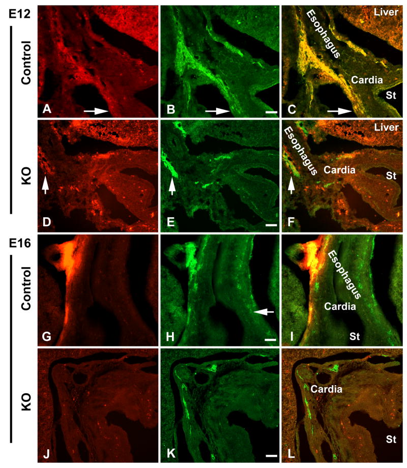
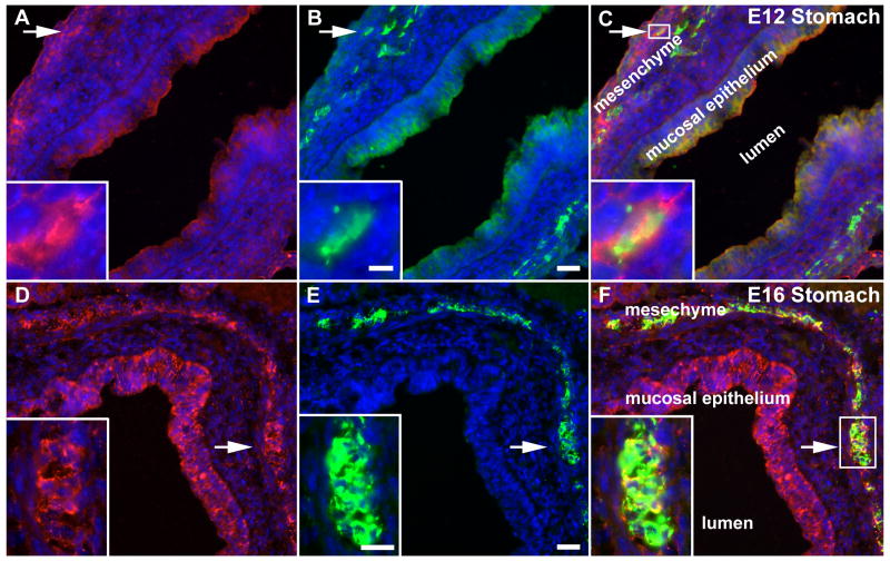
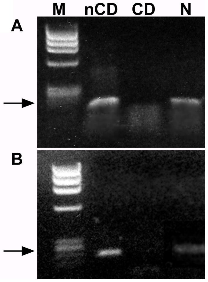
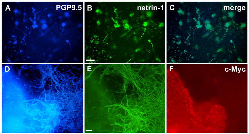
During fetal life, vagal sensory fibers establish a reproducible distribution in the gut that includes an association with myenteric ganglia. Previous work has shown that netrin is expressed in the bowel wall and, by acting on its receptor, deleted in colorectal cancer (DCC), mediates the guidance of vagal sensory axons to the developing gut. Because the highest concentration of netrins in fetal bowel is in the endoderm, we tested the hypothesis that the ingrowth of vagal afferents to the gut would be independent of the presence of enteric neurons, although enteric neurons might influence the internal distribution of these fibers. Surprisingly, experiments indicated that the vagal sensory innervation is intrinsic neuron-dependent. To examine the vagal innervation in the absence of enteric ganglia, fetal Ret -/- mice were labeled by applying DiI bilaterally to nodose ganglia. In Ret -/- mice, DiI-labeled vagal sensory axons descended in paraesophageal trunks as far as the proximal stomach, which contains neurons, but did not enter the aganglionic bowel. To determine whether neurons produce netrins, enteric neural-crest-derived cells (ENCDCs) were immunoselected from E15 rat gut. Transcripts encoding netrin-1 and -3 were not detected in the ENCDCs, but appeared after they had given rise to neurons. When these neurons were cocultured with cells expressing c-Myc-tagged netrin-1, the neurons displayed netrin-1, but not c-Myc, immunoreactivity. Enteric neurons thus synthesize netrins. The extent to which neuronal netrin accounts for the dependence of the vagal sensory innervation on intrinsic neurons, remains to be determined.


Figures

Figure 1
Figure 1
Vagal sensory axons fail to innervate Ret -/- gut. Vagal sensory axons are visualized by applying DiI bilaterally to the nodose ganglia of Ret-sufficient (control) and -deficient (knockout; KO) fetal mice at E12 and E16 (A, D, G, J). Nerves are identified with antibodies to PGP9.5 (B, E, H, K). All images are from coronal-sectioned material; the esophagus, cardia and lumen of the stomach (St) are labeled in the merged images (C, F, I, L). (A) Both paraesophageal bundles and gastric vagal sensory axons are found in the E12 control Ret +/- gut (arrow). (B and C) DiI-labeled vagal axons are double-labeled with antibodies to PGP9.5 (arrow). (D and F) While DiI-labeled vagal axons can be seen in the paraesophageal bundles (arrow), no DiI-labeled gastric vagal sensory axons are found in the E12 Ret -/- stomach. Autofluorescent cells are visualized in the stomach wall. (E) Enteric neurons are absent in the body of the Ret -/- stomach at E12. (G and I) At E16, DiI-labeled vagal fibers can be identified in the wall of the esophagus and adjacent cardiac stomach. (H) Enteric ganglia, visualized with antibodies to PGP9.5, are present in the presumptive myenteric plexus of the Ret +/+ bowel (arrow). (J and L) No DiI-labeled vagal sensory axons are identified distal to the cardiac stomach in the E16 stomach of Ret -/- mice. Autofluorescent cells are visualized in the stomach wall. (K) Enteric nerves, labeled with antibodies to PGP9.5 in the cardiac stomach, are absent in the body of the Ret -/- stomach at E16. Bars = 50 μm (B, E, H, K).
Figure 2
Figure 2
Enteric neurons in the wild-type stomach are netrin-immunoreactive at E12-E16. Stomach from E12 and 16 fetal mice was immunostained to locate netrin-1 (red; A, D) and the neuronal marker, PGP9.5 (green; B, E) in the bowel wall. Nuclei are identified by staining DNA with bisbenzimide (blue). The lumen of the stomach is labeled (lumen) and the images are merged in C and F. (A-C; E12) Netrin-1 immunoreactivity (A, C) is found in the mucosal epithelium and outer mesenchyme (labeled in C). PGP9.5 immunoreactivity (B) overlaps the netrin-immunoreactive band in the outer mesenchyme. At high magnification, a subset of neurons (B; arrow and inset) are also netrin-1-immunoreactive (A, C; arrow, box and inset). (D-F; E16) The gastric mucosal epithelium and outer mesenchyme (labeled in F) are still netrin-1-immunoreactive (D, F). PGP.5 immunoreactivity corresponds in location to the band of netrin-1 immunoreactivity in the outer mesenchyme (E, F). At high magnification, many neurons (E; arrow and inset) are also netrin-1-immunoreactive (E, F; arrow, box and inset). Bars = 25 μm (B, E); 5 μm (B; inset); 15 μm (E; inset).
Figure 3
Figure 3
Enteric neurons, but not their crest-derived precursors, express mRNA encoding netrin-1 and netrin-3. (A) mRNA was isolated from non-crest derived (nCD), crest-derived and from crest-derived cells that had been cultured and differentiated into enteric neurons (N). Transcripts encoding netrin-1 are present in the nCD and N, but not in the CD cells. Arrow = 153 bp. (B) Transcripts encoding netrin-3 are present in the nCD and N, but not in the CD cells. Arrow = 222 bp.
Figure 4
Figure 4
Crest-derived cells, immunoselected from E15 rat gut, were cultured for 6 days in 3-dimensional collagen gels ± c-Myc-tagged netrin-1-secreting 293-EBNA cells. (A) PGP9.5 immunoreactivity (blue). (B) Netrin-1 immunoreactivity (green). (C) Merged image. After 6 days in single culture, the crest-derived cells have differentiated into enteric neurons and are PGP9.5-immunoreactive. (A) The same PGP 9.5-immunoreactive cells are netrin-1-immunoreactive (B, C). (D) PGP9.5 immunoreactivity (blue). (E) Netrin-1 immunoreactivity (green). (F) c-Myc- immunoreactivity (red). After 6 days in culture, PGP9.5-immunoreactive neurons have differentiated and given rise to an exuberant outgrowth of neurites (D). Netrin-1-immunoreactity is coincident with that of PGP9.5 within the neurites (E). Note that the neurites are all free of c-Myc immunoreactivity (F). The plane of focus in panels D and E is through the neurite outgrowth and thus neuronal cell bodies, which lie deep within the collagen gel, are not resolved. Bars = 100 μm (B, E).
Figure 5
Figure 5
Total netrin-1 protein is not decreased in Ret -/- gut. (A) Protein was isolated from 293-EBNA control (C) and netrin-1-secreting cells (N), from E14 mouse brain (Br; n = 3) and from one litter of E14 Ret +/+ (n = 1), Ret +/- (n = 5) and Ret -/- gut (n = 2). The n values refer to numbers of individual mice. Immunoblotting reveals netrin-1 protein in all examples, except for the control cell line negative control. Levels of β-actin protein are similar in all samples. (B) The relative intensity of netrin-1 immunoreactivity was compared to that of β-actin in protein extracted from E14 brain and Ret +/+, Ret +/- and Ret -/- gut. There is no statistical difference in mean relative netrin-1/β-actin intensity between Ret-sufficient (Ret +/+ and Ret +/-) and Ret-deficient (Ret -/-) gut. Dot plot: mean ± SEM.

References

    1. Anderson RB, Stewart AL, Young HM. Phenotypes of neural-crest-derived cells in vagal and sacral pathways. Cell Tissue Res. 2006;323:11–25. - PubMed
    1. Astic L, Pellier-Monnin V, Saucier D, Charrier C, Mehlen P. Expression of netrin-1 and netrin-1 receptor, DCC, in the rat olfactory nerve pathway during development and axonal regeneration. Neuroscience. 2002;109:643–656. - PubMed
    1. Blackshaw LA, Brookes SJ, Grundy D, Schemann M. Sensory transmission in the gastrointestinal tract. Neurogastroenterol Motil. 2007;19:1–19. - PubMed
    1. Chalazonitis A, Rothman TP, Chen J, Lamballe F, Barbacid M, Gershon MD. Neurotrophin-3 induces neural crest-derived cells from fetal rat gut to develop in vitro as neurons or glia. J Neurosci. 1994;14:6571–6584. - PMC - PubMed
    1. Colamarino SA, Tessier-Lavigne M. The axonal chemoattractant netrin-1 is also a chemorepellent for trochlear motor axons. Cell. 1995;81:621–629. - PubMed

Publication types

MeSH terms